
- Title
-
Chemical Screening for Hair Cell Loss and Protection in the Zebrafish Lateral Line
- Authors
- Coffin, A.B., Ou, H., Owens, K.N., Santos, F., Simon, J.A., Rubel, E.W., and Raible, D.W.
- Source
- Full text @ Zebrafish
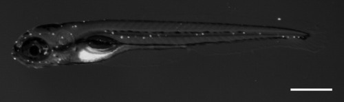
Fluorescent micrograph of a 5 days postfertilization zebrafish labeled with the mitochondrial potentiometric dye DASPEI. Each white dot is a neuromast arrayed along the head and body of the animal. Scale bar = 500 μm. |
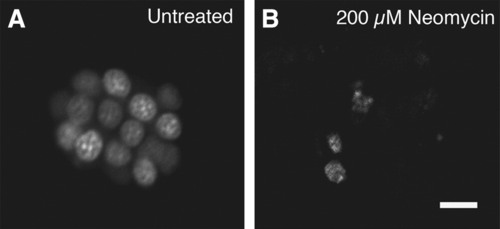
Zebrafish hair cells labeled with the fluorescent dye YO-PRO-1, which binds DNA and labels hair cell nuclei. (A) An undamaged neuromast labeled with YO-PRO-1. Approximately 15 hair cells are visible and healthy in appearance. (B) After a 1-h exposure to the ototoxic drug neomycin at a concentration of 200 μM, most of the hair cells have died. Scale bar in (B)= 10 μm and applies to both panels. Zebrafish hair cells labeled with the fluorescent dye YO-PRO-1, which binds DNA and labels hair cell nuclei. (A) An undamaged neuromast labeled with YO-PRO-1. Approximately 15 hair cells are visible and healthy in appearance. (B) After a 1-h exposure to the ototoxic drug neomycin at a concentration of 200 μM, most of the hair cells have died. Scale bar in (B) = 10 μm and applies to both panels.PHENOTYPE:
|

ZFIN is incorporating published figure images and captions as part of an ongoing project. Figures from some publications have not yet been curated, or are not available for display because of copyright restrictions. PHENOTYPE:
|