FIGURE SUMMARY
Title

Distinct Retinal Pathways Drive Spatial Orientation Behaviors in Zebrafish Navigation

Authors
Burgess, H.A., Schoch, H., and Granato, M.
Source
Full text @ Curr. Biol.

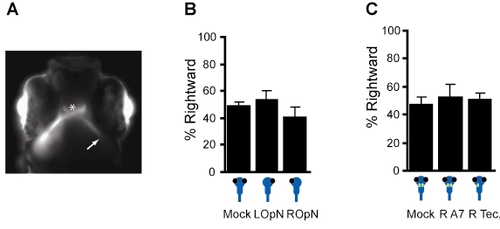
Controls for lesion analysis experiments, related to Figure 4
(A) DiI labeling of retinal ganglion cells after laser ablation of the right optic tectum confirms a loss of the retinotectal projection to the right tectum. Instead, retinal ganglion cell afferents terminate in a stump like bundle (arrow) after crossing at the optic chiasm (asterisk). In contrast, the left optic tectum is clearly labeled. (B) Unilateral section of the optic nerve produces no directional bias in turn movements under uniform illumination (100 μW/cm2). Bars show the percentage of turns initiated in a rightward direction for larvae with mock lesions (′Mock′, n=9 groups), left optic nerve lesions (′LOpN′, n=6 groups) and right optic nerve lesions (′ROpN′, n=5 groups). Similarly, (C) Unilateral laser ablation of the tectum does not produce a bias in turn direction under uniform illumination (40 μW/cm2). ′Mock′ (n=10) larvae were mounted for ablation but not exposed to laser. ′R A7′ (n=11) larvae had visual area rA7 ablated. ′R Tec′ (n=14) larvae had the right optic tectum ablated. Error bars show SE.

Acknowledgments
This image is the copyrighted work of the attributed author or publisher, and ZFIN has permission only to display this image to its users. Additional permissions should be obtained from the applicable author or publisher of the image. Full text @ Curr. Biol.