
- Title
-
Activity-independent specification of synaptic targets in the posterior lateral line of the larval zebrafish
- Authors
- Nagiel, A., Patel, S.H., Andor-Ardó, D., and Hudspeth, A.J.
- Source
- Full text @ Proc. Natl. Acad. Sci. USA
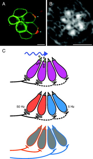
Afferent terminals synapse specifically with hair cells of one orientation. (A) In this anteroposteriorly oriented neuromast of the posterior lateral line in a zebrafish larva at 5 dpf, the axonal terminal of an mCherry-labeled afferent neuron (red) contacts two of the six hair cells marked by GFP (green). Each site of innervation is marked by an arrowhead oriented in the direction of the associated hair cell′s direction of excitatory stimulation. (B) Staining of the same neuromast with fluorescent phalloidin reveals the hair-bundle polarities: the unlabeled kinocilia appear as dark notches in the bright, actin-rich cuticular plates. The two labeled terminals contact hair cells sensitive to anteriorly directed stimuli. Arrowheads mark the hair bundles corresponding to the innervated somata in the two previous panels. In this and all subsequent morphological illustrations, the same labeling convention applies; anterior is to the left and dorsal to the top. The same labeling reagents are used in Figs. 2 and 3. (All scale bars, 5 μm.) (C) Three models might explain the ability of afferent neurons to distinguish between hair-cell polarities. (Top) A posteriorly directed stimulus depolarizes posteriorly polarized hair cells while hyperpolarizing anteriorly polarized cells. Afferents might form synapses diffusely but, after detecting temporal differences in synaptic release from oppositely polarized hair cells, eliminate synapses with hair cells firing out of phase with the rest of their synaptic repertoire (dashed neuronal segment). (Middle) Oppositely polarized hair cells express different complements of ion channels that produce distinct patterns of spontaneous synaptic release. In this example, hair cells of the two orientations release neurotransmitter at different frequencies, allowing neurites to distinguish them. (Bottom) Hair cells express distinct membrane-bound or secreted proteins that attract prepatterned afferents with intrinsic affinities for particular molecular markers. The afferents then detect hair-cell polarities independently of synaptic activity. |
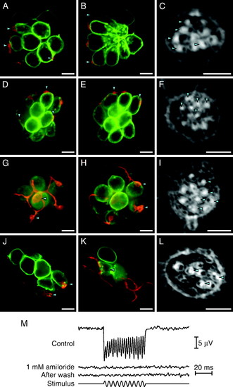
Stimulus-evoked patterns of synaptic release are not required for polarity choice. (A and B) In an anteroposterior neuromast of a tmie mutant larva, a labeled afferent fiber synapses with five of the ten hair cells. In this and the subsequent morphological illustrations, the two micrographs represent different planes of focus. (C) The hair-bundle polarities of this neuromast reveal that the neuron innervates all five posteriorly polarized hair cells and none of the opposite polarity. (D–F) In a dorsoventral neuromast of a tmie mutant, an afferent neuron innervates only the five ventrally polarized hair cells. (G–I) An afferent fiber in a pcdh15a mutant forms synapses with four of the five anteriorly polarized hair cells but with none of the five cells of the opposite polarity. (J–L) In a neuromast of a larva treated with 1 mM amiloride from 2 dpf to 5 dpf, the labeled fiber forms synapses with only the three anteriorly polarized hair cells. (M) The microphonic potential recorded from a neuromast of a 5-dpf larva under control conditions (Top trace) reveals a response at twice the frequency of the 200-Hz, ±8-μm stimulus (Bottom trace). Stimulation of a neuromast from a sibling maintained for 3 days in 1 mM amiloride reveals no microphonic signal (Second trace). Even after extensive washout of the amiloride, the neuromast fails to respond (Third trace). |
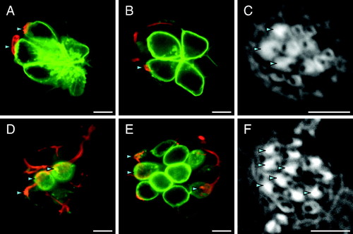
Synaptic transmission is dispensible for hair-cell polarity preference. (A–C) In an anteroposteriorly oriented neuromast of a cav1.3a mutant lacking functional L-type voltage-gated Ca2+ channels, the three mature posteriorly polarized hair cells bear labeled afferent synapses; none of the opposite polarity does. (D–F) This vglut3-deficient neuromast contains six posteriorly polarized hair cells, all of which are innervated by the labeled afferent fiber. |