
- Title
-
Silencing Of Directional Migration In Roundabout4 Knockdown Endothelial Cells
- Authors
- Kaur, S., Samant, G.V., Pramanik, K., Loscombe, P.W., Pendrak, M.L., Roberts, D.D., and Ramchandran, R.
- Source
- Full text @ BMC Cell Biol.
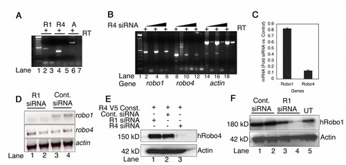
siRNA mediated knockdown of robo4 and robo1. A shows RT-PCR gel for robo1 (R1), robo4 (R4) and actin (A) transcripts in the presence (+) of reverse transcriptase (RT) from HUVEC total RNA. The relative expression level of robo1 has percentile rank of 79 ± 4.5% and robo4 is slightly higher at 83.7 ± 5.7% in HUVECs [35,36]. Numbers on the bottom of gel represent lane numbers. B depicts RT-PCR for robo1, robo4 and actin genes in the presence (+) of reverse transcriptase (RT) from total RNA isolated from HUVECs transfected with increasing concentrations (bars) of robo4 siRNA. For robo1 gene: lanes 4 and 6, robo4 gene: lanes 10 and 12, and actin gene: lanes 16 and 18 represent 125 and 250 ng of siRNA respectively. Lanes 2, 8 and 14 are RNA isolated from untransfected HUVECs, and represent endogenous levels of each transcript. C indicates the ratios of robo1 and robo4 transcripts by real time PCR in control lacZ siRNA and robo4 siRNA transfected cells. D shows the RT-PCR for robo1, robo4, and actin transcripts in robo1 siRNA (lanes 1 and 2) or control lacZ (lanes 3 and 4) siRNA cells. E shows western blots for V5-human Robo4 and actin protein in 293 cell lysates from samples transfected with respective siRNA indicated by +. F shows western blot for endogenous Robo1 and Actin protein levels in control lacZ siRNA (Cont. siRNA) or robo1 (R1 siRNA) and untransfected (UT) 293 cells. Lanes 1 and 3, 2 and 4 are 250 and 500 ng of respective siRNAs indicated on the top of the gel. Lanes 15, 17 and 19 have spillover of excess actin product in -RT lanes. |
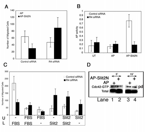
Slit2 mediates chemokinetic and chemotactic behaviour on endothelial cells while serum exclusively mediates chemotaxis. A shows the migration of control lacZ siRNA and robo4 siRNA transfected endothelial cells to AP and AP-Slit2N (25 ng/ml) fusion proteins in a Boyden chamber assay. The data here is consolidated from three independent experiments with each experiment performed with samples in triplicate. B shows the AP activity in lysates prepared from untransfected (UT), AP and AP-Slit2N treated control and robo4 siRNA transfected endothelial cells. C shows migration assay for control lacZ and robo4 siRNA transfected cells to Serum or AP-Slit2N in either upper (U), lower (L) or both chambers as indicated. Error bars in A (n = 3), and B (n = 3) represent SD while in C represent SEM (n = 4). D shows pulldown analysis of Cdc42-GTP levels in AP and AP-Slit2N (25 ng/ml) treated endothelial cell lysates for 5 and 15 minute respectively. + indicate addition of the reagent on the left, pd: pulldown, total: total Cdc42 protein in lysates. |
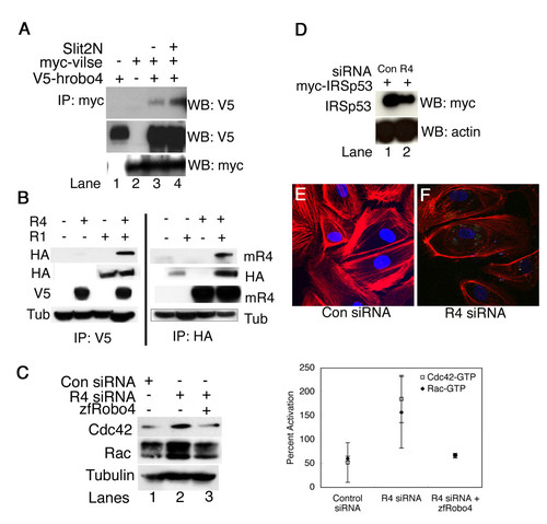
Vilse and Robo1 interact with Robo4 and Rho GTPase homeostasis is important for Robo4 mediated directional migration. A shows the interaction between Robo4 and Vilse in 293 cells. Myc-Vilse and V5-tagged human Robo4 were co-transfected into 293 cells in different combinations as indicated by + and -. Slit2N was added to the transfected cells and lysates were immunoprecipitated by myc antibody followed by western with V5 antibody. B shows IP/Western analysis for rat Robo1 and human Robo4 (left panel) or rat Robo1 and mouse Robo4 (right panel). The antibodies are indicated to the left and right of the gels. Tub: tubulin, mR4: mouse Robo4. The top gel in each panel represents the IP'd lysate western blotted with the indicated antibody. The rest of the gels are western blots for the respective proteins either present (+) or absent (-) as indicated for each sample in the top of the gel. C shows pull down analysis for Cdc42-GTP and Rac-GTP in Con (control) siRNA, robo4 (R4) siRNA and robo4 siRNA plus zfRobo4 transfected endothelial cells. Quantitation of the western blots was performed as described before [22]. Antibodies used for westerns are shown to the left of the blot. D shows western blots with myc and actin antibody of lysates from control lacZ siRNA (lane 1) or robo4 siRNA (lane 2) co-transfected with myc-IRSp53 constructs in endothelial cells. E and F are confocal images of F-actin phalloidin (red) stained endothelial cells transfected with reagents shown below the panel. Slides were mounted in media containing DAPI (blue), which stains nuclei. For A, B, C and D + indicates addition of the reagent on the left, IP-immunoprecipitation, WB-western blot, antibodies indicated on right, For panels C, D, E and F Con: control lacZ siRNA, R4: robo4 siRNA. |