
- Title
-
Proteasomal selection of multiprotein complexes recruited by LIM homeodomain transcription factors
- Authors
- Güngör, C., Taniguchi-Ishigaki, N., Ma, H., Drung, A., Tursun, B., Ostendorff, H.P., Bossenz, M., Becker, C.G., Becker, T., and Bach, I.
- Source
- Full text @ Proc. Natl. Acad. Sci. USA
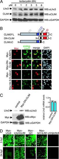
CLIM protects endogenous Lhx3 from proteasomal degradation in αT3 cells. (A) Cellular levels of Lhx3 and CLIM are regulated by the proteasome. Shown are Western blots of protein extracts that were treated for 6 h with increasing amounts of proteasome inhibitor lactacystin. (B) (Upper) CLIM deletion constructs consisting of CLIM2 full length (CLIM2FL; 1–373), dominant-negative CLIM (DN-CLIM; 245–341), and a C-terminal deletion (CLIMΔC; 1–277). DD, dimerization domain; LCCD, Ldb1/Chip conserved domain; NLS, nuclear localization signal; LID, LIM interaction domain. (Lower) Transfection of αT3 cells with Myc-tagged CLIM expression constructs. Cells are costained with specific Myc (red) and Lhx3 (green) antisera. Note the higher levels of endogenous Lhx3 in cells transfected with Myc-CLIM2FL and Myc-DN-CLIM. (C) (Left) Western blot of protein extracts of cells transfected with Myc-DN-CLIM or the empty vector as control. The same blot was probed with antisera against Lhx3, Myc and GAPDH. Note the higher levels of endogenous Lhx3 in cells transfected with Myc-DN-CLIM. (Right) No change in Lhx3 mRNA levels of the same transfected cells as measured by real-time RT-PCR (n = 3; values are mean ± SE). (D) The LIM domain mediates instability of LIM-HD proteins. mRNA encoding LIM-HD proteins Lhx3 and Isl1 with or without LIM domains were injected in one- to two-cell-stage zebrafish embryos. Embryos were fixed at 5 and 24 hpf and stained with a monoclonal Myc antibody. At 24 hpf, the focus is on trunk somites. Note that little or no Myc staining is detected in Myc-Lhx3 and Myc-Isl1-injected embryos, whereas ΔLIM proteins are readily detectable. |
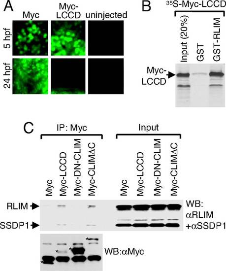
The LCCD region interacts with SSDP1 and RLIM and mediates instability. (A) The LCCD domain functions as an instability domain. mRNA encoding Myc-LCCD was injected in zebrafish embryos. Embryos were fixed at 5 and 24 hpf and stained with a monoclonal Myc antibody. Note that little or no Myc staining is detected in 24 hpf embryos of Myc-LCCD-injected animals, whereas Myc-NLS control proteins are readily detectable. (B) The LCCD region interacts directly with RLIM in vitro. Shown is GST pull-down using GST-RLIM and [35S]Myc-LCCD. (C) The LCCD interacts with RLIM and SSDP1 in cells. Co-IP of SSDP1 and RLIM from αT3 cells transfected with Myc-LCCD. (Upper) Western blot of Myc IPs (Left) and 100% input control (Right) using antibodies directed against RLIM and SSDP1 (10% gel). (Lower) To visualize Myc-proteins, an aliquot of the same Myc-IPs was run on a 15% gel in parallel. Myc proteins are indicated by asterisks. Lower Myc-LCCD and Myc-CLIMΔC protein levels probably reflect their instability in cells. Bands not marked with asterisks are unspecific. |