
- Title
-
Hox cofactors in vertebrate development
- Authors
- Moens, C.B., and Selleri, L.
- Source
- Full text @ Dev. Biol.
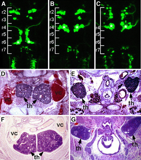
Pbx mutants in the fish and mouse recapitulate Hox mutant phenotypes. (A–C) Facial motor neuron migration defects in hox and pbx mutants in the zebrafish. (A) Wild-type zebrafish carrying an isl1-GFP transgene that is expressed in cranial motor neurons. Note that the facial motor neurons have migrated to r6 (asterisks), leaving the facial nerve behind to exit from r4. (B) isl1-GFP transgenic injected with an antisense morpholino (MO) that blocks the function of Hoxb1a. Note that the facial motor neurons have differentiated in r4 (asterisk) but have not undergone their posterior migration. (C) isl1-GFP transgenic zebrafish lacking zygotic Pbx4 protein due to a genetic mutation. Note that the facial motor neurons have failed to migrate (asterisk). (D–G) Thymic defects in Hox and Pbx mutants in the mouse. Transverse H&E-stained sections through E16 thymic lobes are shown. (D, F) Wild-type littermates of the mutants shown in panel E, G. (E) Pbx1-/- embryo; (G): Hoxa3+/-; Hoxb3-/-; Hoxd3-/- compound mutant. In both mouse mutants, the thymic phenotype is identical: the lobes are ectopic (still localized within the neck), they do not fuse and do not descend into the mediastinum as in wild-type embryos. Thymus, th; superior vena cava, vc (lower panels are adapted from Manley and Capecchi, 1998). |
Reprinted from Developmental Biology, 291(2), Moens, C.B., and Selleri, L., Hox cofactors in vertebrate development, 193-206, Copyright (2006) with permission from Elsevier. Full text @ Dev. Biol.