
- Title
-
Churchill regulates cell movement and mesoderm specification by repressing Nodal signaling
- Authors
- Londin, E.R., Mentzer, L., and Sirotkin, H.I.
- Source
- Full text @ BMC Dev. Biol.
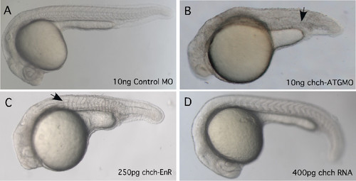
chch inhibition results in axial and somite defects. The effect of inhibiting chch was examined using a morpholino and a dominant-negative construct. Microinjection of chch-ATGMO results in a broad, misshapen notochord (arrow) and misshapen somites and a shortened axis (B). Microinjection of chch-EnR, results in a similar phenotype as the morpholino, a broad, misshapen notochord, and enlarged and misshapen somites formed (C). chch mRNA overexpression produces embryos that are wild-type in appearance (D). Arrowheads point to the notochord. All embryos are 24 hpf. PHENOTYPE:
|
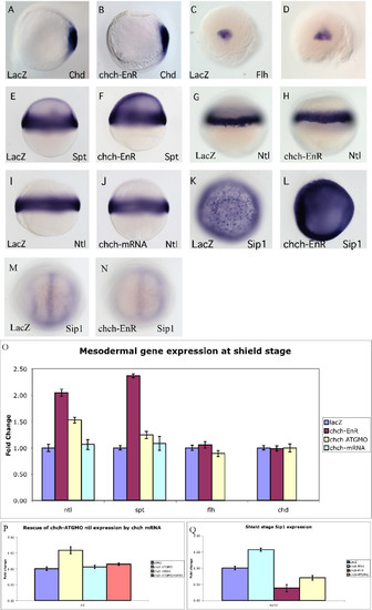
chch represses mesodermal markers. Mesodermal markers were examined by RNA in situ hybridization and real-time PCR in embryos microinjected with chch-EnR mRNA. Inhibition of chch does not alter expression of dorsal mesodermal genes chd (A, B) or flh (C, D). Expression of mesodermal markers at shield stage, including spt (E-F) and ntl (G-H) are expanded slightly in chch-inhibited embryos. Overexpression of chch mRNA does not alter no-tail expression at shield stage (I-J). chch inhibition results in a decrease in Sip1 gene expression at bud stage (M-N), while chch activation results in Sip1 induction at shield stage (K-L). Real-time PCR analysis of mesodermal markers in chch-inhibited embryos (O). The fold change in transcript levels (y-axis) is graphed relative to control embryos following overexpression of chch-EnR mRNA, chch-ATGMO and chch mRNA. This analysiinhibited embryos (O). The fold change in transcript levels (y-axis) is graphed relative to control embryos following overexpression of EXPRESSION / LABELING:
|
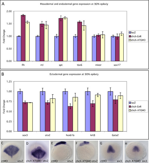
chch inhibition results in decreased ectodermal gene expression. Real-time PCR analysis of mesodermal, ectodermal and endodermal markers during late gastrulation in chch-compromised embryos. The mesodermal markers flh, ntl, spt, and tbx6 (A), endodermal markers, mixer and sox17b (A) and ectodermal markers otx2, hoxb1b, sox3, krt8 and gata2 (B) were examined following microinjection of chch-EnR mRNA and chch-ATGMO. Mesodermal markers are increased, endodermal are unaffected while ectodermal marker levels are decreased. Expression of the neural genes otx2 at 90% epiboly (C-D) and 6-somites (E-F) and Six3 at 6-somites (G-H). C-D, G-H are dorsal views. E-F are lateral views. EXPRESSION / LABELING:
|
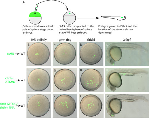
chch inhibition results in inappropriate cell movements. (A) A schematic representation of the transplantation scheme. ctMO or chch-ATGMO mRNA-injected donor cells were transplanted to the hemisphere of a sphere stage wild-type host. Embryos were photographed at 40% epiboly (B, F, J), germ ring (C, G, K) shield stage (D, H, L), and 24 hpf (E, I, M). The chch morphant cells (F-I) undergo greater spreading than the control cells and move toward the margin. At 24 hpf control cells are often found in anterior neural tissue (E) while the chch morphant cells are more frequently found in superficial cell layers of the trunk (I). The abnormal movement of chch morphant cells to superficial cell layers of the trunk is rescued by the injection of chch-mRNA (J-M). |
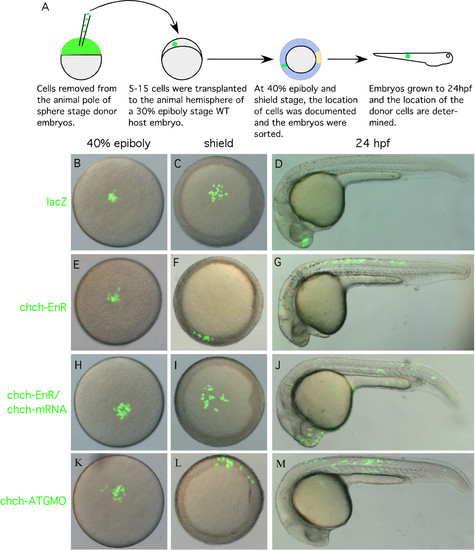
chch-inhibited cells leave the epiblast and enter the germ ring and become mesoderm. (A) A schematic representation of the transplantation scheme. Cells from sphere stage LacZ, chch-EnR mRNA or chch-ATGMO injected donors were transplanted to the animal hemisphere of 30% epiboly (late blastula) wild-type embryos. Embryos were photographed at 40% epiboly (B, E, H, K), at the start of gastrulation (shield) (C, F, I, L) and after 24 hpf (D, G, J, M). As expected, LacZ cells remain within the ectoderm and take on ectodermal fates at 24 hpf (B-D). In contrast, chch inhibited cells often migrate from the presumptive ectoderm to the germ ring (E-G, K-M). These cells are often later found in the somites (G). The abnormal movement of chch-EnR cells into the germ ring is rescue by coinjection of chch mRNA (H-J). The images in each row are of the same embryo. |
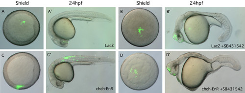
Alk receptor signaling is required for both migration and acquisition of mesodermal character of chch-EnR transplanted cells. Transplanted LacZ cells were present in the animal pole at shield stage (A) and were found in anterior neural tissue after 24 hpf (A′). 400 µM SB431542 (Sigma-Aldrich), which inhibits Alk receptors did not alter LacZ donor cell behavior (B-B′). Conversely, chch-EnR donor cells moved from the animal hemisphere to the germ ring by shield stage (C) and were observed in mesodermal structures after 24 hpf (C′). SB431542 treatment results in blocking movement of chch-EnR cells. The donor cells remain in the animal hemisphere at shield stage (D) and are found in anterior neural structures after 24 hpf (D′). This result suggests that signaling via Alk receptors is required for both migration and acquisition of mesodermal character of chch-EnR donor cells. The images in each row are of the same embryo. |
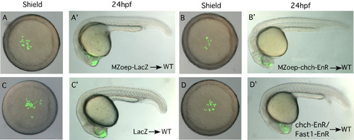
chch-compromised cells migrate to the germ ring and become mesoderm in response to Nodal signals. Transplanted MZoep-LacZ cells to the animal hemisphere of a wild-type host remain within the animal hemisphere at shield stage (A) and are observed in anterior neural structures after 24 hpf (A'). Similar behavior was observed for transplanted MZoep-chch-EnR cells in wild-type hosts (B-B'). Similarly, when donor cells express 12.5 pg xFast1-EnR, along with chch-EnR, migration to the margin is blocked and donor cells are observed in anterior neural structures after 24 hpf (D-D'). Together, these results demonstrate that the migration of the chch inhibited cells to the germ ring and acquisition of mesodermal character depends upon Nodal signaling. |