
- Title
-
A hierarchy of Runx transcription factors modulate the onset of chondrogenesis in craniofacial endochondral bones in zebrafish
- Authors
- Flores, M.V., Lam, E.Y., Crosier, P., and Crosier, K.
- Source
- Full text @ Dev. Dyn.
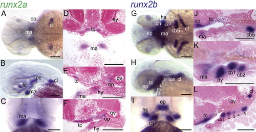
Comparison of the expression patterns of duplicated zebrafish runx2 genes in the developing pharyngeal arches by whole-mount in situ hybridization of 2dpf embryos with runx2a (A-F) and runx2b (G-L) riboprobes. (A,G) Dorsal and (B,H) lateral views, anterior to left; (C,I) ventral view, anterior to top; (D) frontal section, anterior to top; (E,F,J-L) sagittal sections, anterior to left. cb, ceratobranchial; ch, ceratohyal; cl, cleithra; ep, ethmoid plate; hy, hyoid; hs, hyosymplectic; ma, mandibular; op, opercula; ov, otic vesicle; pc, parachordal; tc, trabecula cranii. Scale bars = 100 μm. EXPRESSION / LABELING:
|
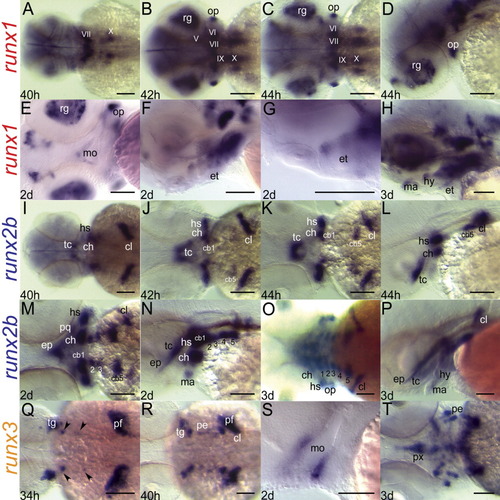
Expression patterns of the runx genes at the onset of and during craniofacial cartilage development (34hpf to 3dpf). (A-H) runx1; (I-P) runx2b; (Q-T) runx3; (A-C, I-K, M,O,Q,R,T) dorsal view; (D,F,H,L,N,P) lateral view left; (E) ventral view; (G,S) magnified lateral view, oral region. All embryos anterior to left. Roman numerals V-X denote expression of runx1 in the cranial nerve ganglia. Presumptive retinal ganglion cells (rg) of the eye and the opercula are also labelled with runx1 riboprobe. Transcripts for runx2b are prevalent in the pre-cartilage mesenchyme, cleithra, and opercula, while those for runx3 mark the trigeminal ganglia, the pharyngeal endoderm, and the pectoral fin anlagen. Early runx3 expression in the pouch endoderm (black arrowheads) is positioned just posterior to the trigeminal ganglia. cb, ceratobranchial (numbers denote cb identity); ch, ceratohyal; cl, cleithrum; ep, ethmoid plate; et, epithelium; hy, hyoid; hs, hyosymplectic; ma, mandibular; mo, mouth; op, operculum; ov, otic vesicle; pc, parachordal; pe, pouch endoderm; pf, pectoral fin; pq, palatoquadrate; px, pharynx; tc, trabecula cranii. Scale bars = 100 μm. EXPRESSION / LABELING:
|
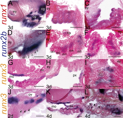
Analysis of tissue sections confirms compartmentalized localization of runx transcripts in the pharyngeal domain. (A-C) runx1; (D-F) runx2b; (G-L) runx3; (A,B,G,J-L) sagittal sections, anterior to left; (B,E,H) frontal sections, anterior to left; (C,F,I) frontal sections, anterior to top. A sagittal section through a 3dpf embryo reveals runx1 expression in pharyngeal arches (A). Frontal sections verify staining in the entire epithelial layer of the pharyngeal arches (B,C). runx2b expression is restricted to mesenchymal cells deep in the core of the arches (D-F), while runx3 marks the endodermal epithelium (G-I) in 3dpf larvae. This endodermal expression of runx3 is present at 2dpf in the pouch endoderm (J), and later in the endodermal lining of the mouth and pharynx (K, L). At 4dpf, runx3 is expressed in differentiating chondrocytes (arrowheads, L). cb, ceratobranchial (numbers denote cb identity); cg, cranial nerve ganglia; hs, hyosymplectic; mo, mouth; px, pharynx; ov, otic vesicle; v, blood vessel. Scale bars = 50 μm. |
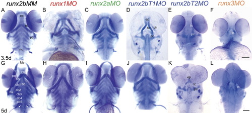
Loss-of-function analyses using antisense morpholinos demonstrate which Runx proteins are essential at the onset of endochondral bone formation in the head skeleton. Embryos were injected with optimal amounts of MO at the 1-4 cell stage to inhibit gene function, and cartilage development was visualized with alcian blue staining at 3.5 (A-F) and 5 (G-L) dpf. (A,G) runx2bMM; (B,H) runx1MO; (C,I) runx2aMO; (D,J) runx2bT1MO; (E,K) runx2bT2MO; (F,L) runx3MO. Compared to runx2bMM controls, embryos treated with runx2bT1MO, runx2bT2MO, and runx3MO display significant reductions in chondrocyte differentiation at 3.5dpf (D-F). By 5dpf, runx2T1 morphants recover from an early delay in chondrogenesis (J), but those injected with runx2bt2MO and runx3MO show persistence of cartilaginous abnormalities with underdeveloped and reduced cartilage (K,L). The head skeleton of runx1 and runx2a morphants are mildly misshapen at 3.5dpf (B,C), and these become morphologically normal by 5dpf (H,I). Scale bars = 100 μm. PHENOTYPE:
|
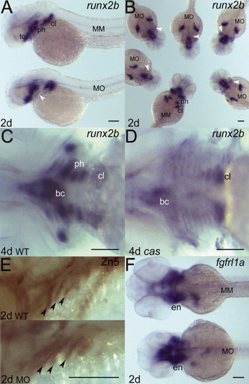
Morpholino knock-down of Runx3 does not influence pharyngeal endoderm formation, but abolishes runx2b expression in the pharyngeal arch mesenchyme. Whole-mount in situ hybridization with a runx2b riboprobe on 2dpf embryos injected with runx3MO reveals specific loss of runx2b expression in the pre-cartilage condensations of pharyngeal arches in runx3 morphants (white arrowheads), while retaining expression in the basicranial anlagen (A,B). Pharyngeal cartilage runx2b expression in 4dpf wild-type larvae (C) is also absent in casanova mutants that lack endoderm (D). However, pharyngeal endoderm is intact in runx3 morphants, as highlighted by immunostaining with Zn5 (E), and normal expression of pouch endoderm marker fgfrl1 (F) in 2dpf embryos. (A,C) Lateral and (B) dorsal views, anterior to left; (D) dorsal view of a group of morphants (MO) and control (MM) embryos. bc, basicranium; cl, cleithrum; en; endoderm; ph, pharyngeal arch cartilage; tc, trabecula cranii. Scale bars = 100 μm. |