
- Title
-
A β1,4-galactosyltransferase is required for Bmp2-dependent patterning of the dorsoventral axis during zebrafish embryogenesis
- Authors
- Machingo, Q.J., Fritz, A., and Shur, B.D.
- Source
- Full text @ Development
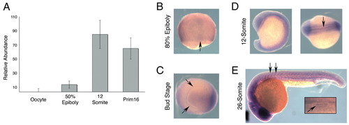
Temporal and spatial expression of ß4galt5 during zebrafish embryogenesis. (A) RT-PCR analysis of staged RNA provides a temporal profile of ß4galt5 expression. There was no detectable level of ß4GalT5 in oocytes. Expression was first detected in the early gastrula embryo (50% epiboly), and reached a steady state by mid-somitogenesis. Error bars indicate s.e.m. (B-E) Whole-mount in situ hybridization of ß4galt5. Expression is widespread throughout the embryo with some refinement in stage-specific structures. (B) Dorsal view of 80% epiboly embryo. Note expression throughout the embryo with a slight increase in the dorsal axis (arrow); peripheral stain reflects `edge effects' due to oocyte curvature. (C,D) Near ubiquitous expression throughout the bud (C, anterior view) and 12-somite stage embryo (D, lateral view, dorsal view at hindbrain level). Note expression in the developing polster (arrows, C) and floorplate (arrow, D). (E) Twenty-six-somite stage embryos (lateral view). Arrows indicate higher expression in intersomitic boundary. Inset illustrates expression at the midline of the otic vesicle (arrow). |
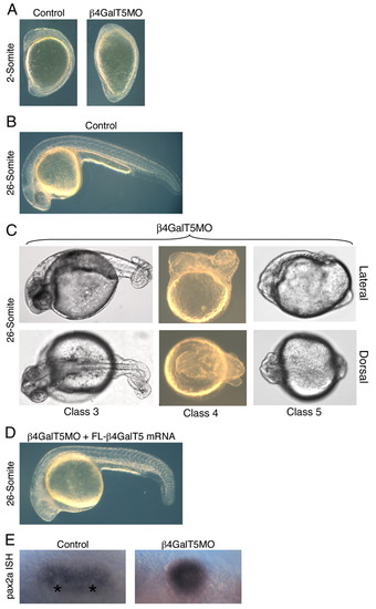
Knockdown of ß4GalT5 results in dorsalization. (A) Lateral views of 2-somite control-injected and ß4GalT5MO-injected embryos. The morphant phenotype is manifested by an elongation of the anteroposterior axis. (B) 26-somite embryo injected with control morpholinos, lateral view. (C) When ß4GalT5MO-injected embryos reach the 26-somite stage, the mature ß4GalT5MO phenotype is observed (lateral and dorsal views). (Class 3) 5 ng of ß4GalT5 morpholino results in mild dorsalization manifested by a slight tail coil. This phenotype is similar to the pgy phenotype reported by Mullins et al. (Mullins et al., 1996) and correlates with their Class 3. (Class 4) 10 ng of morpholino produces a more significant coiling of the tail, as well as dorsalization within the anterior regions of the embryo, and embryos are considered moderately dorsalized, similar to the snh phenotype representing Class 4 of Mullins et al. (Mullins et al., 1996). (Class 5) Injection of 15-20 ng of ß4GalT5 morpholino produces the most severe dorsalization, which appears similar to that seen in the swr mutant. (D) Lateral view of 26-somite embryo injected with ß4GalT5MO3 and mRNA encoding full-length ß4GalT5. These embryos were essentially wild type in appearance. (E) In situ hybridization of pax2a in the otic vesicle of 26-somite control embryo; asterisks indicate the paired otoliths, which are absent in an equivalently staged ß4GalT5MO embryo. EXPRESSION / LABELING:
|
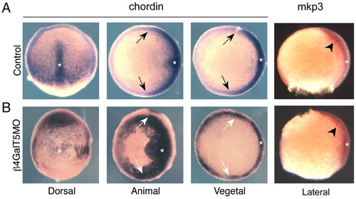
Misexpression of chordin in ß4GalT5MO embryos. (A) In situ hybridizations of chordin in control-injected embryos show that expression is restricted to the dorsal axis (asterisk in all panels). Arrows indicate the limits of chordin expression and identify the presumptive dorsoventral boundary. (B) Embryos injected with ß4GalT5MO display disorganized chordin expression, including invasion into the presumptive ventral domain. For comparison, the limits of chordin expression in control embryos (A) are indicated by the white arrows. In ß4GalT5 morphants, chordin expression extends beyond the boundaries seen in control embryos and envelope the ventral hemisphere. In contrast to that seen with chordin, the expression of the Fgf target mkp3 (arrowheads) appears unaffected in morpholino-injected embryos. EXPRESSION / LABELING:
|
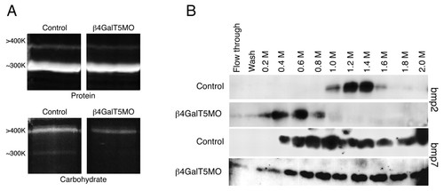
Decreased binding of BMP2 to ß4GalT5MO proteoglycans. (A) Proteoglycans isolated from control- and ß4GalT5MO-injected embryos were resolved by 6% SDS-PAGE into two high molecular weight bands. Protein and carbohydrate content were revealed by staining with SPYRO and Pro-Q Emerald, respectively. Protein levels in each molecular weight species appear similar between control and ß4GalT5MO samples, whereas the extent of glycosylation is dramatically reduced in ß4GalT5MO embryos. (B) Proteoglycans from control-injected or ß4GalT5MO-injected embryos were bound to an affinity support and assayed for their ability to bind recombinant BMP2 and BMP7. BMP2 showed peak elution from control proteoglycans at 0.8-1.6 M NaCl. BMP2 eluted from ß4GalT5MO proteoglycans at 0.2-0.8 M NaCl. Interestingly, there was no significant difference in the ability of control or ß4GalT5MO proteoglycans to bind recombinant BMP7. Similar results were obtained using proteoglycans isolated from embryos injected with either splice-blocking (MO2) or translation-blocking (MO3) oligonucleotides. |