
- Title
-
Requirement for intracellular calcium modulation in zebrafish dorsal-ventral patterning
- Authors
- Westfall, T.A., Hjertos, B., and Slusarski, D.C.
- Source
- Full text @ Dev. Biol.
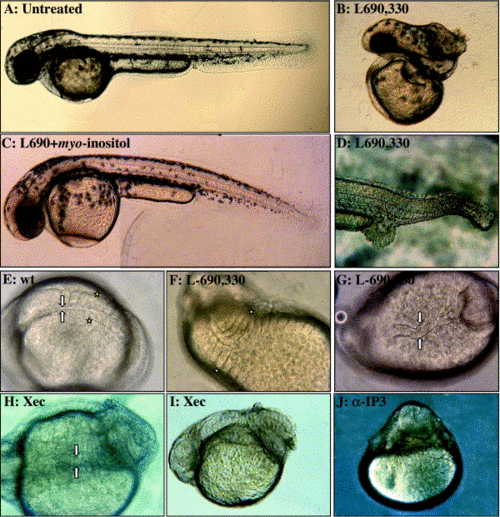
Range of morphological phenotypes induced by PI cycle inhibition. Lateral view with anterior to the right of live embryos approximately 48 hpf (A–D). (A) Untreated. (B) L-690,330-injected (3–6 mM) with reduced tail tissue (similar to lost-a-fin, C2). (C) control L-690,330 (10 mM) + myo-inositol-injected (50 mM). (D) L-690,330-injected (10 mM, snail-house-like, C4). Dorsal (E–G) and lateral (I–J) view of ∼24 hpf embryos. (E) Wild-type embryo with arrows denoting notochord width and stars highlight the lateral portion of one pair of somites. L-690,330-injected embryos display both (F) expanded somites, stars and (G) thickened notochord, arrows similar to swirl-like phenotypes (C5). Embryos treated with IP3Receptor inhibitors display (H) thickened, wavy notochord (arrows, XeC). (I) tail curling phenotypes similar to piggy-tail-like (C3) (XeC), and (J) monoclonal blocking antibody-induced bustling and radialization of tissue over the yolk. |

Phenotypes generated from PLC Inhibition. Lateral view of live embryos at 24 hpf after incubation in (A) Control (U-73343) and (B). Inhibitor (U-73122) at 5x magnification. Lateral posterioer view of the tail after 36 hpf of (C). Control (U-73343) and (D) Inhibitor (U-73122)-injected embryos at 10x magnification. (E) Embryo coinjected with U-73122 and myo-inositol. Anterior is to the left in all panels. |

Window of sensitivity and axis duplication induced by PI cycle inhibition. Lateral view with anterior to the left of live embryos after 48 hpf. Late treatment of zebrafish embryos with PI cycle inhibitors results in anterior head defects and fusion of the eyes (U-73122, 2.4%; XeC, 3.2%; and L-690,330, 5.7%). (A) Control embryo with star denoting one eye in focus. (B) Embryo injected at a late stage with L-690,330 with the star denoting a reduced eye with partial cyclopia. PI cycle inhibition generates duplicated dorsal structures. (C) L690,330-injected embryo with duplicated heads (arrows). The fourth eye is slightly out of the focal plane (white arrow). (D) XeC-injected embryo with the partial secondary axis including ectopic ears (white arrow) and abnormal pericardial cavity. The primary axis folded over the yolk (black arrow). |

Expanded and ectopic chordin domains in PI-inhibited embryos. Expression pattern of chordin at 50% epiboly in PI-inhibited embryos. Animal pole view with dorsal to the right (A–D). (A) Wild-type embryo, arrows denote the lateral domain of chordin expression. Embryo treated with (B) IP3R inhibitor (XeC) with arrows showing expanded chordin domain. (C) L-690,330-injected and (D) thapsigargin-treated embryos with radialized chordin expression. Dorsal view of (E) Control (10 mM L-690,330 + 50 mM myo-inositol-injected) embryo with a relatively normal chordin expression domain (stars) and of (F) L-690,330-injected embryo with an ectopic domain, arrow. |

PI cycle inhibition increases the extent of nuclei containing β-catenin at sphere stage. Immunolocalization of β-catenin protein in sphere-stage embryos. Four-micron optical sections were collected with confocal microscopy. Embryos were oriented with the animal pole up. (A, D, and I) 20x magnification; (B, C, E–H, J–M) 63x magnification. The approximate locations of the higher magnification images are mapped, in lower case, onto the 20x image. (A) Wild-type embryo at sphere stage, 20x representation for orientation. Representative 63x images of the same embryo collected around the circumference are shown in (B) and (C) with dots noting the location of nuclear β-catenin. β-Catenin-positive nuclei are localized to a region of approximately one-third the circumference of the embryo. (D) XeC-injected embryo at 20x for orientation with individual high magnification images are shown in (E–H), dots noting intense nuclear β-catenin localization. XeC-treated embryos have expanded nuclear β-catenin domains that span more than 50% the circumference of the embryo. The L-690,330-injected embryo in (I) has nuclear β-catenin in ectopic locations that span the circumference of the embryo. Clusters of β-catenin-positive nuclei are present in (L) and (M) (L, M) β-Catenin localization at region opposite the location of (J) and (K). Numerous internal cells with nuclear β-catenin are not marked with dots. |

Ectopic domains of bozozok in PI-inhibited embryos. Whole-mount in situ hybridization of bozozok at sphere-stage embryos following PI cycle inhibition. Dorsal view of (A) untreated with stars noting the endogenous boz domain and (B). XeC-injected embryo with a robust primary boz expression domain (stars) and ectopic domains (arrow). Animal pole views of (C) control embryo, stars noting the endogenous expression and (D) L-690,330-treated with the radialization of boz expression highlighted by a dashed line. |
Reprinted from Developmental Biology, 259(2), Westfall, T.A., Hjertos, B., and Slusarski, D.C., Requirement for intracellular calcium modulation in zebrafish dorsal-ventral patterning, 380-391, Copyright (2003) with permission from Elsevier. Full text @ Dev. Biol.