
- Title
-
Gelsolin is a dorsalizing factor in zebrafish
- Authors
- Kanungo, J., Kozmik, Z., Swamynathan, S.K., and Piatigorsky, J.
- Source
- Full text @ Proc. Natl. Acad. Sci. USA
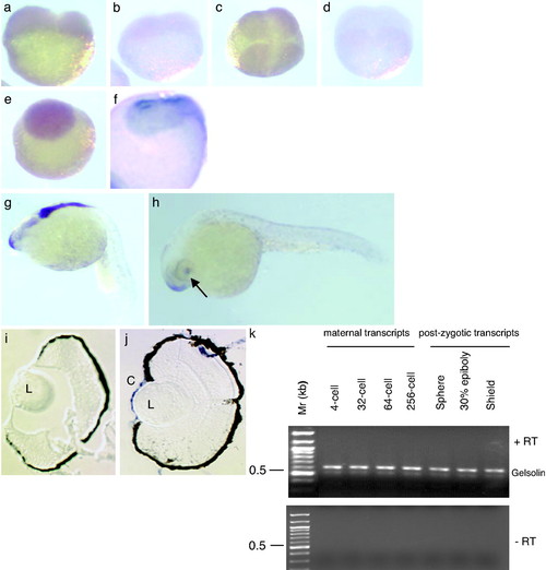
Developmental expression of gelsolin mRNA. Gelsolin mRNA expression is shown in four stages of embryo by using an antisense probe: two-cell (a), four-cell (c), blastula (e), and late-gastrula (f). A sense probe was used to hybridize in the two-cell (b) and four-cell (d) embryos. The gastrula (e) is photographed, magnified x2 to show the anterior part of the embryo. Gelsolin expression in the forebrain and hindbrain is shown in a 22-h-old embryo (g), in the snout and cornea of a hatching-stage embryo (h), and in the sections of eyes from a hatching-stage embryo probed with the sense (i) and antisense (j) riboprobes. Arrow indicates the location of the eye. Locations of the lens (L) and cornea (C) are indicated. (k) RT-PCR for gelsolin by using cDNA synthesized from embryonic RNA obtained from the indicated developmental stages. (Upper) Reaction products with reverse transcriptase (+RT). (Lower) Products without the enzyme (–RT). EXPRESSION / LABELING:
|

MO knockdown of gelsolin ventralizes embryos. Embryos 30 hpf after microinjection of control MO (2.5 ng/E; a) and gelsolin MO (b) are shown. Arrows indicate the location of the eyes. Embryos injected with control (c) or gelsolin MO (1 ng/E; d) are shown 72 hpf. Gelsolin MO resulted in ventralized embryos (b) and hatchlings with smaller and less pigmented eyes (d). The effect of different doses of gelsolin MO on the gelsolin and actin levels was determined by immunoblotting of embryo extracts prepared 8 h after MO injection (e; ref. 14). A representative immunoblot of four separate experiments is shown. The phenotypic effect of different doses of the gelsolin MO is summarized (f). Histological analyses using hematoxylin/eosin staining compare the normal and reduced development of anterior cephalic structures in control (g) and gelsolin MO-injected (h) embryos at 30 hpf. Locations of brain (B), eye (E), and yolk mass (Y) are indicated. Gelsolin mRNA (50 pg/E) injection results in dorsalized embryos (i and j). Arrows indicate duplicated posterior axial regions. Microinjection of human plasma gelsolin protein (4 ng/E) resulted in axis duplication (k). Arrows indicate duplicated anterior axial region including head and eyes. Histological section (l) of the embryo with duplicated axes showing notochord (nc) and optic cup (oc). Coinjection of gelsolin MO (2.5 ng/E) with 4 ng of human gelsolin protein (m), or with gelsolin mRNA (50 pg/E; n), or with chordin mRNA (50 pg/E; o) rescued the gelsolin MO-injected phenotypes. Embryos at 30 hpf are shown. PHENOTYPE:
|
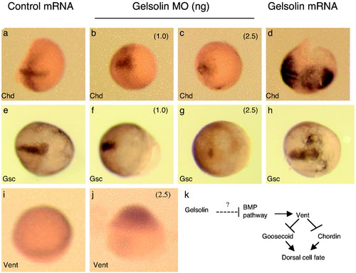
In situ hybridization demonstrates the expression of chordin (a–d), goosecoid (e–h), and Vent (i and j) in embryos harvested 8 h postinjection with control mRNA synthesized from PCS2 vector control or gelsolin mRNA, as indicated. Control (a and e), gelsolin MO-injected (1 ng/E; b and f), gelsolin MO-injected (2.5 ng/E; c and g), and gelsolin mRNA-injected (50 pg/E; d and h) embryos are shown, with the anterior side to the right. Vent mRNA expression is shown in embryos injected with control MO (i) and gelsolin MO (j) at 2.5 ng/E. The animal pole of the embryos is oriented upward. (k) A schematic of how gelsolin might regulate cell fates in zebrafish. |