
- Title
-
Chondroitin sulfates affect the formation of the segmental motor nerves in zebrafish embryos
- Authors
- Bernhardt, R.R. and Schachner, M.
- Source
- Full text @ Dev. Biol.
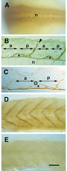
The distribution of chondroitin sulfate immunoreactivity in the zebrafish embryo and its elimination by chondroitinase treatment are shown by immunostaining using mAb 473. (A, D, and E) Whole-mount preparations; (B and C) semithin sections. Anterior is to the left, in D and E dorsal is to the top. (A) A low-power dorsal view of an 18-hpf embryo shows accumulation of chondroitin sulfate labeling in the posterior half of each somite, adjacent to the notochord (n). The segmental myosepta are also labeled. (B) A horizontal section through a 22-hpf embryo reveals chondroitin sulfate reactivity around the notochord (asterisk), around putative migrating sclerotome cells (arrows), and in the segmental myosepta (arrowhead). (C) A high-power view of a horizontal section through a 24-hpf embryo shows the relationship of the ventral motor nerve (labeled by the tubulin antibody, circled) to putative migrating cells and chondroitin sulfate immunoreactivity which appears relatively faint in this doubly labeled preparation. A comparison with B will confirm, however, that the migrating cells along which the axons grow are surrounded by chondroitin sulfates. (D) A side view of a 26-hpf embryo shows the prominent chondroitin sulfate reactivity in the segmental myosepta. (E) Similar view of a corresponding embryo injected with chondroitinase ABC at 16 hpf. No chondroitin sulfate reactivity can be detected. Magnification bar: 100 μm for (A), 25 μm for (B), 10 μm for (C), and 70 μm for (D and E). |
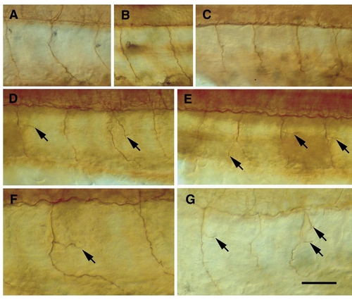
Ventral motor nerves are revealed by immunostaining for acetylated α-tubulin in control (A–C) and in experimental embryos (D–G). Lateral views of whole-mount preparations, anterior is to the left, dorsal to the top. Arrows indicate abnormal side branches of the ventral motor nerve. (A) Ventral motor nerves (somites 7–9, left) extend without side branches along a midsegmental pathway in an uninjected embryo (26 hpf). (B) Anti-tubulin staining does not reveal side branches at least up to 36 hpf in uninjected embryos (shown here are ventral motor nerves 8 and 9, left). (C) Ventral motor nerves (7–10, right) are unbranched in an embryo (26 hpf) injected with vehicle solution. (D) Abnormal side branches have formed in two ventral motor nerves (8 and 10, left) in an embryo (26 hpf) injected with chondroitinase ABC (Sigma). (E) Same embryo as in (D), abnormal side branches have formed in three ventral motor nerves (8 –10, right). (F) Abnormal side branch in a ventral motor nerve (10, left) in an embryo (26 hpf) injected with chondroitinase ABC (Sigma). (G) Abnormal side branches in two ventral nerves (9 and 11, right) in an embryo (26 hpf) injected with chondroitinase ABC (Sigma). Magnification bar: 50 μm for (A–E), 30 μm for (F and G). |
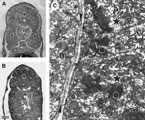
Light and electron micrographs of cross sections through two 22-hpf embryos injected with chondroitinase ABC (Sigma; B, C), and through an uninjected embryo (A). In the injected embryos chondroitin sulfates are expected to be missing even if their absence was not confirmed by immunostaining in order to ensure good ultrastructural preservation. (A) A semithin section through an uninjected embryo stained with toluidine blue. s, spinal cord; n, notochord; asterisks, somite. (B) A semithin section stained with toluidine blue shows no evidence of nonspecific damage in the spinal cord, notochord, and somite (labeled as in A). The box indicates the approximate frame of the electron micrograph shown in (C) that illustrates another embryo. (C) Electron micrograph of the interface of somite and notochord at the level of the horizontal myoseptum. The microenvironment through which the ventral motor nerve extends shows no evidence of structural damage. In particular the basal lamina (between arrowheads) that separates the notochord from the somite (asterisks) appears intact, as does the medial surface of the somite (between arrows). Within the somite muscle cells have differentiated normally as indicated by the presence of actin filament clusters (a). Magnification bar: 25 μm for (A and B), 3 μm for (C). |
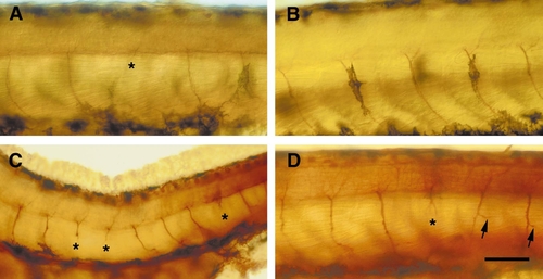
Ventral motor nerves are revealed by immunostaining for acetylated α-tubulin in embryos injected with different chondroitin sulfates. Lateral views of whole-mount preparations, anterior is to the left, dorsal to the top. Embryos are 32–34 hpf. Asterisks indicate truncated or missing ventral motor nerves, arrows abnormal side branches. (A) Injection of the chondroitin sulfate mixture (25 mg/ml) has led to truncated ventral motor nerves, illustrated are segments 12–16 on the right body side. (B) Injection of chondroitin sulfate-C (25 mg/ml) has not affected the ventral motor nerves, illustrated are segments 10–15 on the right body side. (C) Injection of chondroitin sulfate-B (25 mg/ml) has induced truncated ventral motor nerves, illustrated in this overview are segments 8–16 on the left body side. (D) Injection of chondroitin sulfate-B (5 mg/ml) has induced truncated and branched ventral motor nerves, illustrated are segments 11–16. Magnification bar: 50 μm for (A, B, D), 100 μm for (C). |
Reprinted from Developmental Biology, 221(1), Bernhardt, R.R. and Schachner, M., Chondroitin sulfates affect the formation of the segmental motor nerves in zebrafish embryos, 206-219, Copyright (2000) with permission from Elsevier. Full text @ Dev. Biol.