
Fig 3
- ID
- ZDB-IMAGE-200220-23
- Publication
- Messerli et al., 2020 - Adaptation mechanism of the adult zebrafish respiratory organ to endurance training
- All Figures
- Figures for Messerli et al., 2020
|
Fig 3
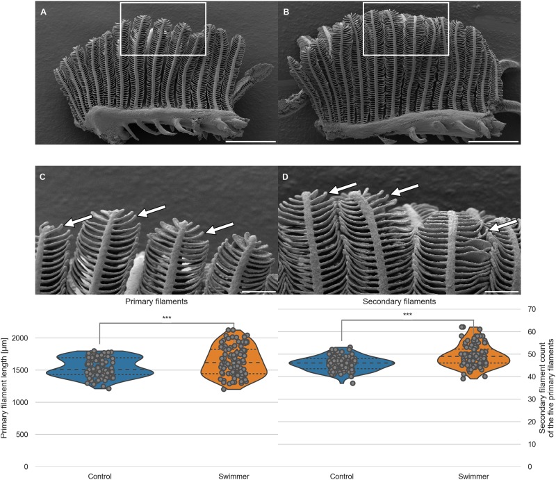
A, B: Gill arch of control (A) and swimmer fish (B) with primary filaments pointing up vertically, secondary filaments are seen on each side of the primary filaments (examples shown with arrows). Also visible are the gill rakers, facing towards the pharynx and preventing food particles from exiting between the gill arches. After 5 weeks of training, SEM scans were printed and compared morphologically. The white frame marks the region of the zoom shown in panel C and D. Scale bars: 0.5 mm. C, D: Detailed view of the tips of the gill arches. Note the longer appearing secondary (arrows) of filaments of the swimmer (D) and their more horizontal appearance. Scale bars: 0.1 mm. Bottom row: Graphs from semi-quantitative measurement of primary filament length and number of secondary filaments on primary filaments, controls and swimmers after the training period. Arches 2 and 3 of each fish were taken into account (n = 100 per group). Left: The length of the five longest primary filaments increased significantly in trained fish (p = 0.00043). Right: secondary filament count on the 5 longest primary filaments of controls and swimmers, the swimmers showed a significantly higher number of secondary filaments (p = 9e-9). ***: p<0.001, lines within the plots show the quartiles of the respective distributions.