
|
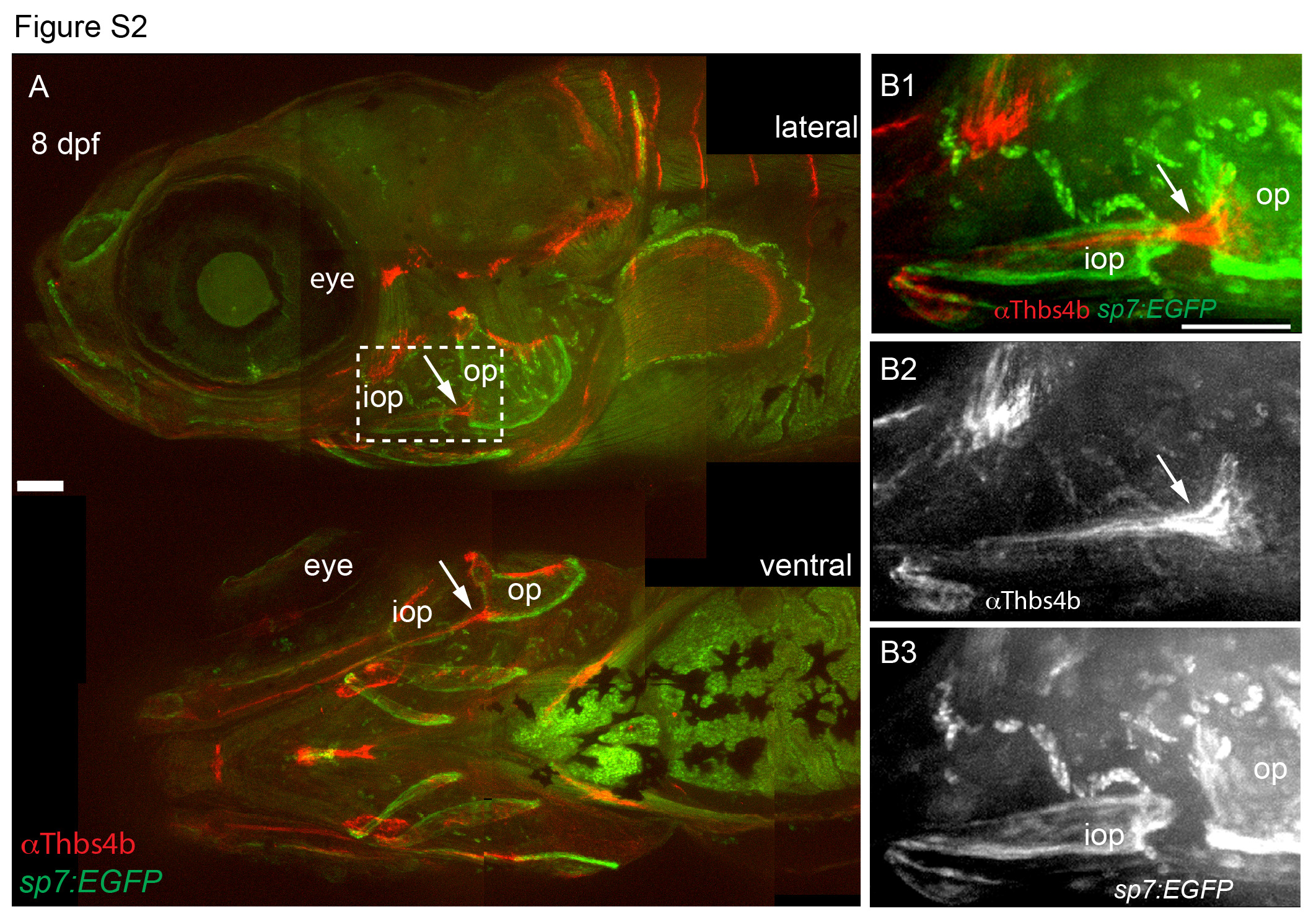
Fig. S2
The interopercle bone develops around the operculohyoid ligament, anterior to the opercle. (A) 8 days post fertilization (dpf) sp7:EGFP wild-type larvae that began feeding at 4 dpf were fixed and stained with anti-Thbs4b and imaged by confocal microscopy. Multiple confocal projections were assembled to make a composite image. The dashed region is enlarged in B. (B) Enlargements of boxed region in A, including single channel fluorescent images of ligaments and tendons in B2 and osteoblasts in B3. The operculohyoid ligament is marked with an arrow, the opercle (op) and interopercle (iop) bony elements are indicated and teh eye is shown for reference. The late developing nature of the iop precludes analysis of this bone in mef2cab1086 mutants. All images are confocal projections and lateral views where anterior is towards the left and dorsal is upward. Scale bars are 100 µm.