
|
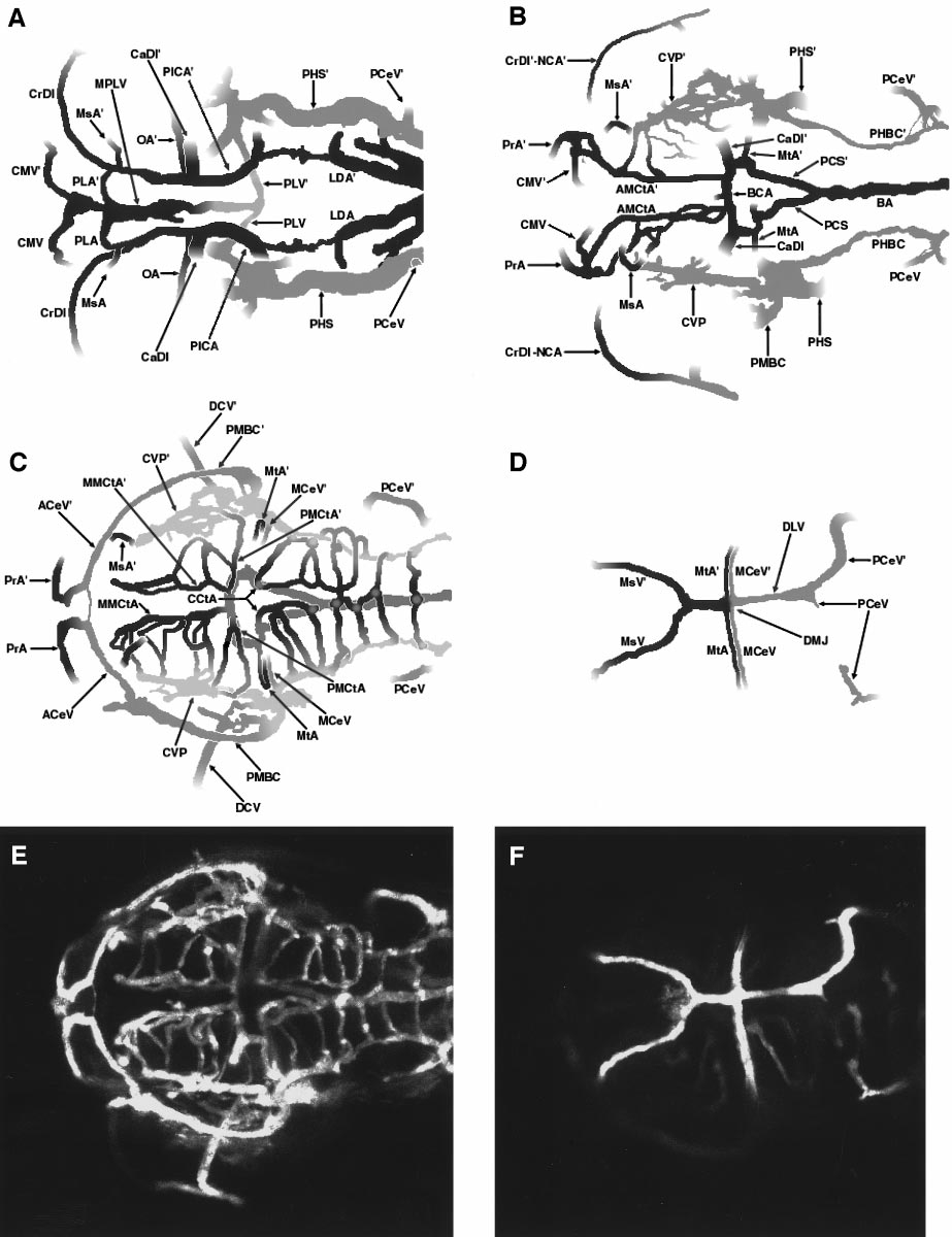
Fig. 5 Multilayer composite diagram of circulation in the developing zebrafish head at approximately 2.5 days postfertilization (dpf). A 2.5 dpf dorsal–view angiographic image stack was divided into 4 substacks and each substack was separately reconstructed. Drawings were prepared detailing the vascular patterns in each of these reconstructions. (A) Bottom layer diagram, showing ventral cranial vessels (beginning approximately just above the future pharynx). (B) Lower middle layer diagram. (C) Upper middle layer diagram. (D) Top layer diagram, showing the most dorsal cranial vessels. (E) Angiogram corresponding to the diagram in panel (C). (F) Angiogram corresponding to the diagram in panel (D). All panels are oriented with rostral to the left and left down. A glossary of the names corresponding to all labeled vessels is provided in Table 1.
Reprinted from Developmental Biology, 230(2), Isogai, S., Horiguchi, M., and Weinstein, B.M., The vascular anatomy of the developing zebrafish: an atlas of embryonic and early larval development, 278-301, Copyright (2001) with permission from Elsevier. Full text @ Dev. Biol.