Image


                        Figure Caption


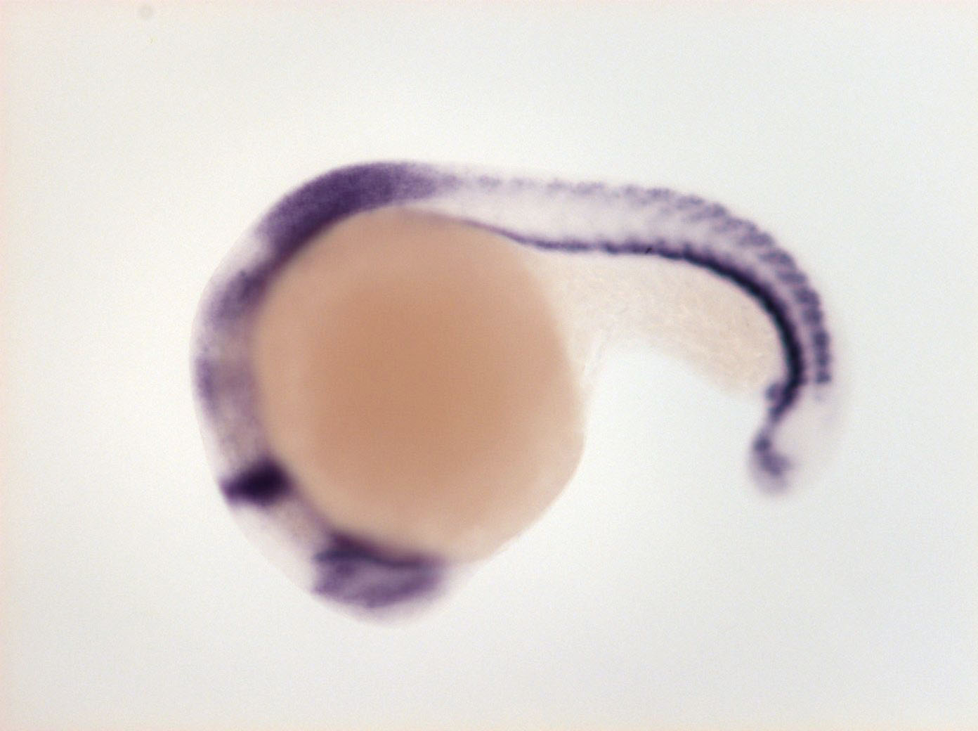
Fig. 3 retina, diencephalon-mesencephalon border, MHB, migrating neural crest cells, rhombomere 3 and 5, anterior spinal chord, somites (ventral and dorsal part), tail bud, ventral mesenchyme
                
                    
                        Orientation


                | Preparation | Image Form | View | Direction | 
| whole-mount | still | side view | anterior to left | 
                
                    
                        Figure Data


        