
Image
|
Figure Caption
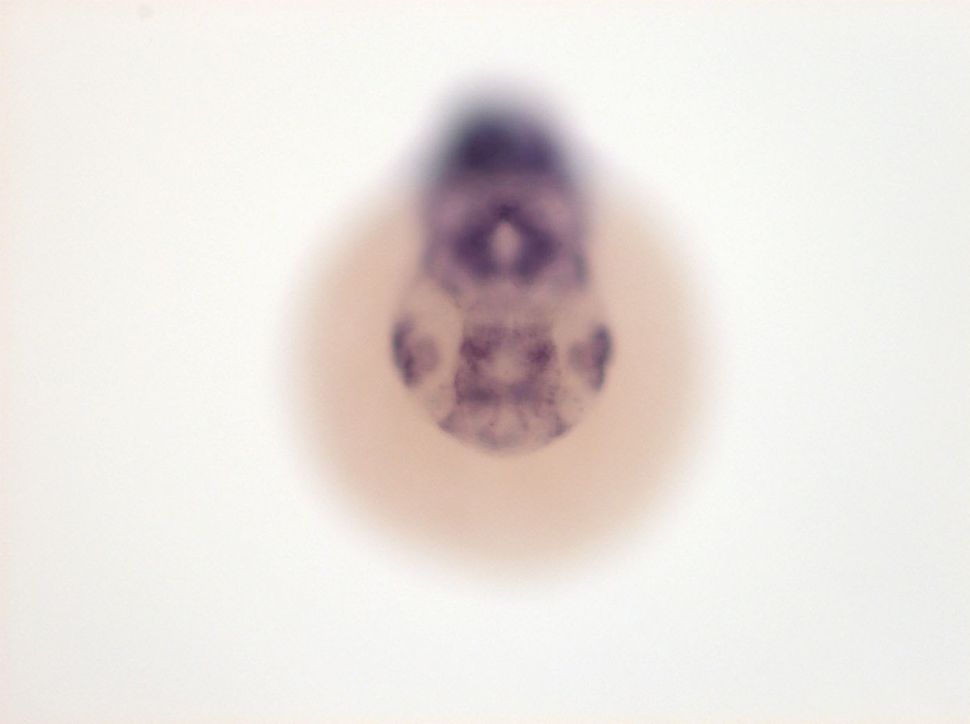
Fig. 4 ventral retina, lens, anterior diencephalon, anterior most ventral diencephalon, MHB +++, hindbrain rhombomeres, pharyngeal arches, anterior spinal chord, somites, hypophysis
Orientation
| Preparation | Image Form | View | Direction |
| whole-mount | still | frontal | anterior to top |
Figure Data