
|
Image description by: Tanya Whitfield
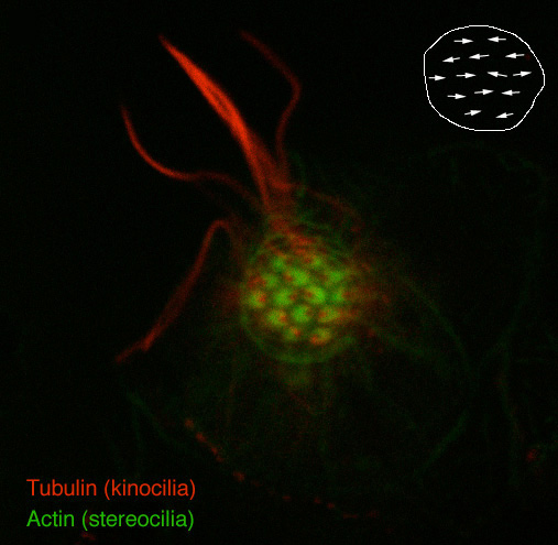
Anatomical structures shown: stereocilia and kinocilia of the hair cells of a lateral line neuromast
Stage: day 5
Genetic (background) strain: none given
Genotype: phenotypically wild-type embryo from a dogtp85b/+ x dogtp85b/+ cross
Animal state: fixed
Labeling: FITC-phalloidin (green), anti-acetylated tubulin antibody (red)
Description: Polarity patterns in a lateral line neuromast. View of a neuromast end on; fluorescein-phalloidin labels the hair cell stereocilia, seen here as green crescents, and anti-acetylated tubulin stains the kinocilium as a red dot on the concave side of each crescent. The inset diagrams the polarities of these hair cells, which lie in two opposing directions. In the trunk, hair cells are aligned predominantly anteroposteriorly. In the head, neuromasts may have anteroposterior or dorsoventral polarities. The entire length of some of the kinocilia can be seen; the curvature of these is an artefact of fixation. In the live animal they would all be projecting straight out of the neuromast into a gelatinous cupula.
Publication containing this image: T. Whitfield (unpublished).
| Preparation | Image Form | View | Direction |
| whole-mount | still | parasagittal | anterior to left |