
|
Image description by: Tanya Whitfield
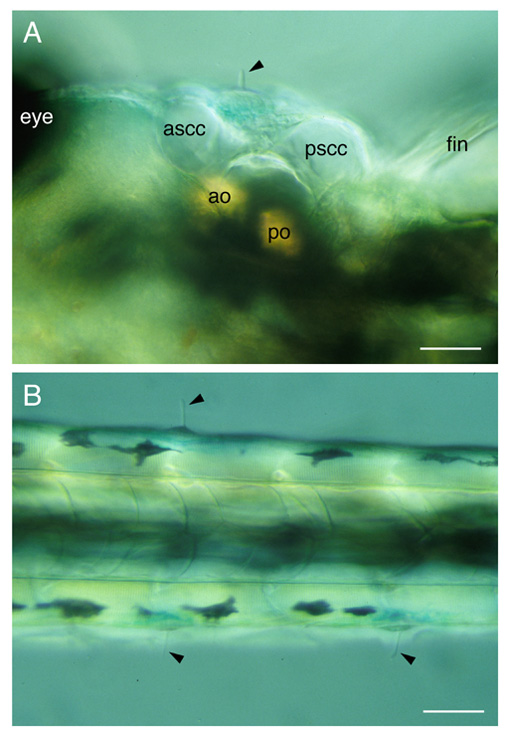
Anatomical structures shown: lateral line neuromasts, cupulae, ear
Stage: day 4
Genetic (background) strain: WIK
Genotype: wild-type
Animal state: live
Labeling: none
Description: Orientation: dorsal, anterior to left. Appearance of lateral line neuromasts in a live day 4 larva. Under the dissecting microscope, neuromasts appear as small bumps decorating the body surface. Under the compound microscope, the gelatinous cupula projecting out of each neuromast can be seen with DIC optics (arrowheads). This contains the long kinocilia of the neuromast hair cells. A. Head region. The MI1 neuromast is obvious over the ear. ascc, pscc, anterior and posterior semicircular canals (seen in cross section); ao, po, anterior and posterior otoliths. B. Trunk. Scale bar, 50 µm.
Publication containing this image: T. Whitfield (unpublished).
| Preparation | Image Form | View | Direction |
| whole-mount | still | not specified | not specified |