
|
Image description by: Tanya Whitfield
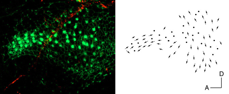
Anatomical structures shown: hair cell polarity patterns in the day 5 posterior macula
Stage: 5 d
Genetic (background) strain: none given
Genotype: wild-type
Animal state: fixed
Labeling: FITC-phalloidin (green); anti-acetylated tubulin (red)
Description: Polarity patterns of hair cells in a day 5 posterior macula. At this stage there are two regions of different orientation. In the slim anterior projection, hair cells are arranged in an antiparallel fashion, with dorsally-located hair cells pointing posteriorly and ventrally-located hair cells pointing anteriorly. In the rounded posterior region, hair cells point away from a midline separating dorsal and ventral halves; dorsal cells point dorsally and ventral cells point ventrally.
Publication containing this image: Adapted from Haddon et al., 1999.
| Preparation | Image Form | View | Direction |
| whole-mount | still | parasagittal | anterior to left |