
- Title
-
Neuroendocrinology of reproduction: Is Gonadotropin-Releasing Hormone (GnRH) dispensable?
- Authors
- Whitlock, K., Postlethwait, J., Ewer, J.
- Source
- Full text @ Front. Neuroendocrinol.
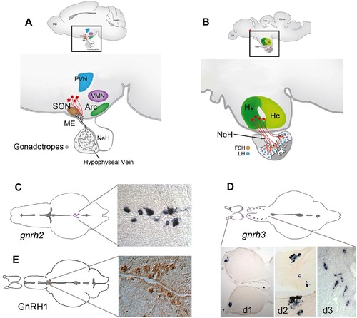
Structure of the vertebrate hypothalamus and description of gnrh/GnRH containing cells in the adult zebrafish brain. (A, B) Schematic diagram of a lateral view of the mouse (A) and the zebrafish (B) brain. In mammals (A), the GnRH cells (red) project to the median eminence, a highly vascularized connection to the pituitary. In teleosts (B), the neurohypophysis (NeH) is reduced and the GnRH cells make neural connections with the pituitary. (C-E) gnrh2 and gnrh3 expression detected by in situ hybridization in the midbrain (C), and terminal nerve (D, d1, d2) and ventral telencephalon (D, d3) (Whitlock, 2005) In contrast, GnRH cells in the parvocellular nucleus of the hypothalamus can be detected only by immunohistochemistry (E from Cortes-Campos et al., 2015). There are no convincing data supporting cellular expression of any gnrhgene in the parvocellular nucleus of the adult zebrafish. Abbreviations: Arc, arcuate nucleus; Hv, ventral zone of periventricular hypothalamus; Hc, caudal zone of periventricular hypothalamus; (red asterisk), neurosecretory preoptic area; ME, median eminence; NeH, neurohypophysis; OB, olfactory bulb; PVN, paraventricular nucleus; SON, supraoptic nucleus; TeO, tectum opticum; VMN, ventromedial nucleus. |