
- Title
-
Motor Neuron Abnormalities Correlate with Impaired Movement in Zebrafish that Express Mutant Superoxide Dismutase 1
- Authors
- Robinson, K.J., Yuan, K.C., Don, E.K., Hogan, A.L., Winnick, C.G., Tym, M.C., Lucas, C.W., Shahheydari, H., Watchon, M., Blair, I.P., Atkin, J.D., Nicholson, G.A., Cole, N.J., Laird, A.S.
- Source
- Full text @ Zebrafish
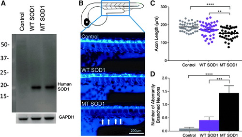
(A) Expression of human SOD1 protein in zebrafish embryos injected with WT or MT (A4V) human SOD1 was confirmed via Western blot analysis (human SOD1 detected at ∼20 kDa). GADPH was used as a loading control (37 kDa). (B) Representative images of the first five spinal motor neurons after the zebrafish yolk sac are shown for zebrafish larvae at 48 hours postfertilization. Control (noninjected) and larvae that expressed WT human SOD1 displayed long, J-shaped motor axons, while those that expressed MT SOD1 had axons that were shorter in length (arrows). (C) Motor neuron axon length analysis revealed larvae that expressed MT SOD1 had significantly shorter axons than noninjected controls (****p < 0.001) or larvae that expressed WT SOD1 (**p = 0.004). There was no statistically significant difference in motor axon length between controls and larvae that expressed WT SOD1. (D)Larvae that expressed MT SOD1 had significantly more aberrantly branched axons per embryo than noninjected controls (****p < 0.001) or those expressing WT SOD1 (***p = 0.001). Each dot represents an individual larva; noninjected: n = 40; WT SOD1: n = 44; MT SOD1: n = 37. WT, wild type; MT, mutant; SOD1, superoxide dismutase 1. |