
- Title
-
A novel early onset phenotype in a zebrafish model of merosin deficient congenital muscular dystrophy
- Authors
- Smith, S.J., Wang, J.C., Gupta, V.A., Dowling, J.J.
- Source
- Full text @ PLoS One
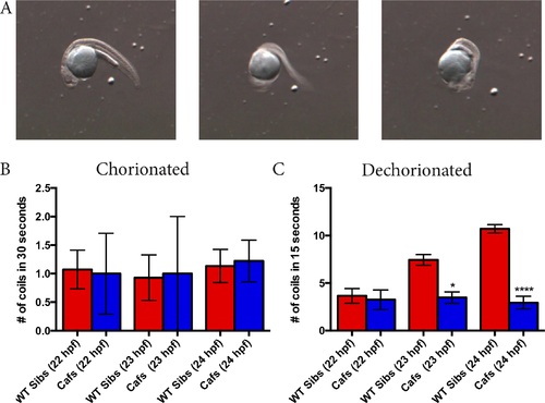
Caf mutants display a coiling phenotype as early as 23 hpf. A) One full coil completed by a 24 hpf wild type (lama2+/+ and lama2+/-; WT) zebrafish embryo. B) Number of coils of WT siblings and caf (lama2-/-) mutants over 30 seconds when left in their chorions. There is no significant difference in the coiling abilities of caf mutants and their WT siblings at 22 hpf (WT sibs: 1.071 ± 0.3391, n = 14; cafs: 1.000 ± 0.7071, n = 4; p>0.9999), 23 hpf (WT sibs: 0.9286 ±0.3987, n = 14; cafs: 1.000 ± 1.000, n = 2; p>0.9999) or 24 hpf (WT sibs: 1.133 ± 0.2906, n = 15; cafs: 1.222 ± 0.3643, n = 9; p>0.9999). C) Number of coils of WT siblings and caf mutants in the 15 seconds immediately after manual dechorionation. At 22 hpf, there was no significant difference in number of coils completed by WT siblings and caf mutants (WT sibs: 3.654 ± 0.7685, n = 26; cafs: 3.250 ± 1.031, n = 8; p>0.9999). However, cafs completed significantly less coils than their WT sibs at 23 hpf (WT sibs: 7.444 ± 0.5587, n = 72; cafs: 3.476 ± 0.5921, n = 21; p = 0.0194) and 24 hpf (WT sibs: 10.71 ± 0.4436, n = 163; cafs: 2.936 ± 0.6646, n = 47; p<0.0001). Bars represent mean ± SEM. PHENOTYPE:
|
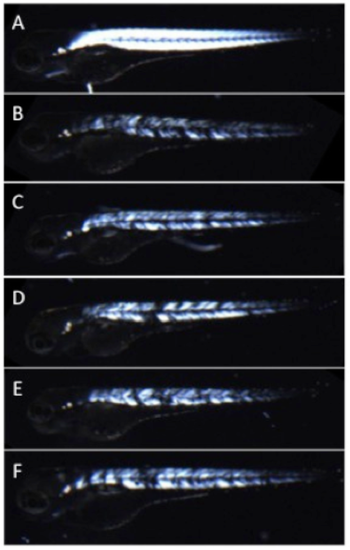
Muscle fiber detachment of caf mutants can be observed with birefringence. Under plane polarized light, muscle from WT siblings (A) appears uniformly bright and white, consistent with normal muscle organization. In contrast, Caf mutants (B-F_ can be identified as having stochastic patterns of muscle degeneration and detachment with birefringence as early as 2 dpf. This is seen in the muscle compartment as dim white areas (thinned or atrophied fibers) and black spots (presumed areas of muscle fiber detachment). Of note, genotype for all depicted animals was confirmed by Sanger sequencing. Bars represent mean ± SEM. PHENOTYPE:
|

ZFIN is incorporating published figure images and captions as part of an ongoing project. Figures from some publications have not yet been curated, or are not available for display because of copyright restrictions. PHENOTYPE:
|

Abnormal phalloidin staining in skeletal muscle from caf mutant embryos. Wild type (WT) and caf mutants were staining with phalloidin to illuminate filamentous actin and then visualized whole mount by confocal microscopy. (A) WTs show the expected pattern of staining at 24 hpf (n = 5). (B) In muscle from caf mutants, there is an accumulation of intense staining in the region of the myotendinous junction (arrow) (n = 5). Scale bar = 10 um. PHENOTYPE:
|
|
ZFIN is incorporating published figure images and captions as part of an ongoing project. Figures from some publications have not yet been curated, or are not available for display because of copyright restrictions. PHENOTYPE:
|
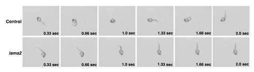
lama2cl501 mutants have reduced coiling upon dechorionation Still images from time lapse videos of wild type clutchmates (control) and lama2cl501 (lama2) mutants at 24 hours post fertilization. Videos were taken just after dechorionation. Lama2 mutants demonstrate only partial coiling and do not complete a normal/full coiling in the 2 second period shown. In contrast, the control embryo completes 2 full coils. PHENOTYPE:
|