- Title
-
Differential activation of natriuretic peptide receptors modulates cardiomyocyte proliferation during development
- Authors
- Becker, J.R., Chatterjee, S., Robinson, T.Y., Bennett, J.S., Panáková, D., Galindo, C.L., Zhong, L., Shin, J.T., Coy, S.M., Kelly, A.E., Roden, D.M., Lim, C.C., and MacRae, C.A.
- Source
- Full text @ Development
|
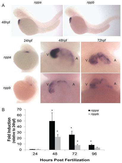
Developmental induction of cardiac natriuretic peptides peaks at 48 hpf in the embryonic zebrafish. (A) Whole-mount in situ hybridization of nppa and nppb zebrafish embryos. A, atrium; V, ventricle. (B) Quantitative RT-PCR measurement of nppa and nppb during different developmental time points. Data are expressed as mean + s.e.m. *P<0.05, relative to 24 hpf values. |
|
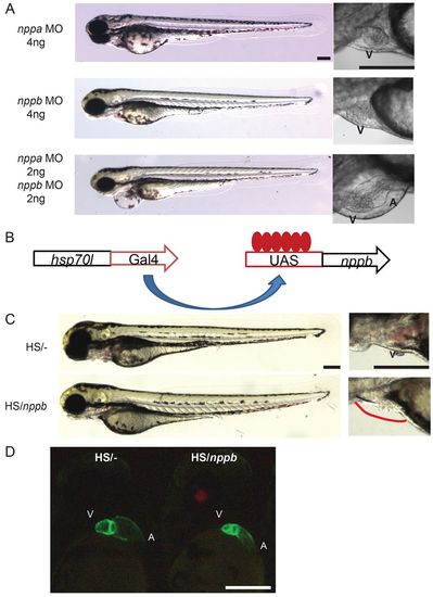
Altered cardiac natriuretic expression changes heart growth in vivo. (A) Morpholino knockdown of Nppa or Nppb in isolation or simultaneously. Heart enlargement was seen in the Nppa/Nppb double knockdown embryos but was absent in single knockdown morphants (72 hpf). (B) Inducible Gal4/UAS transactivator system to overexpress nppb in vivo. (C) The nppb overexpression embryos (HS/nppb) had a large reduction of pericardial fluid volume (magnified panel, red line details normal pericardial border) but overall embryonic growth was minimally altered (72 hpf). (D) The cmlc2/CFP fluorescent marker was utilized to visualize the chamber dimensions better in control (HS/-) and nppb overexpression (HS/nppb) embryos (48 hpf). The ventricular and atrial sizes were both reduced in nppb overexpression embryos. Red fluorescent marker in lens denotes UAS/nppb carrier status. A, atrium; V, ventricle. Scale bars: 200 μm. EXPRESSION / LABELING:
PHENOTYPE:
|
|
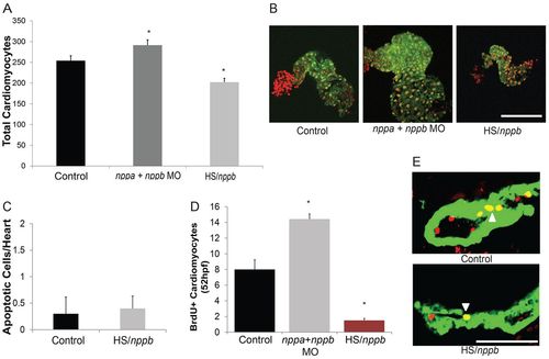
Manipulation of cardiac natriuretic peptide expression alters total cardiomyocyte numbers in the embryonic heart. (A) Total cardiomyocytes at 48 hpf were increased in the Nppa/Nppb double knockdown (nppa/nppb MO) embryos, but were decreased in the nppb overexpression embryos (HS/nppb). *P<0.02 compared with control. (B) Representative images from cmlc2:CFP (pseudo-colored green) and DAPI (pseudo-colored red) stained hearts. (C) The total number of apoptotic heart cells was not increased in the nppb overexpression embryos. P>0.05 compared with control. (D) Knockdown of Nppa/Nppb and overexpression of nppb were able to significantly alter cardiomyocyte proliferation at 52 hpf. *P<0.03 compared with control. (E) Representative images of BrdU-labeled (red) cmlc2:CFP (green)-positive heart tissue. White arrowhead indicates BrdU-positive cardiomyocytes. Intracavitary BrdU+ cells were considered to be endothelial in origin and were not counted. All data are expressed as mean + s.e.m. Scale bars: 100 μm. |
|
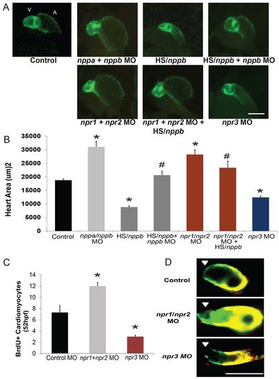
The cardiomyocyte proliferation effects of the cardiac natriuretic peptides are coordinated by the action of multiple natriuretic peptide receptors in vivo. (A) Lateral views of 48 hpf cmlc2:CFP embryos show significantly altered atrial and ventricular chamber sizes in Nppa/Nppb double knockdown and nppb overexpression embryos. Likewise, double knockdown of both natriuretic peptide guanylate cyclase-linked receptors, Npr1 and Npr2, caused a similar phenotype as the Nppa/Nppb double knockdown and could block the effect of nppb overexpression (npr1/npr2 MO + HS/nppb). Finally, the knockdown of Npr3 caused a significant reduction in both atrial and ventricular chamber sizes that was similar to that observed after overexpression of nppb. (B) Corresponding heart surface areas of all treatment groups shown in A. *P<0.05 compared with control heart size, #P<0.05 compared with HS/nppb alone. (C) Cardiomyocyte proliferation was increased in the Npr1 and Npr2 knockdown embryos, whereas Npr3 knockdown embryos had reduced cardiomyocyte proliferation at 52 hpf as assessed by BrdU labeling of cardiomyocytes. *P<0.01 compared with control. (D) Differentiation of ventricular cardiomyocytes between 30 and 48 hours of heart development was not altered by knockdown of the natriuretic peptide receptors. White arrowhead denotes arterial pole, areas of green tissue were added after 30 hpf (non-photoconverted). Yellow tissue is a mixture of photoconverted red tissue and non-photoconverted green tissue. All data are expressed as mean + s.e.m. Scale bars: 100 μm. |


