
- Title
-
APOBEC2, a selective inhibitor of TGFβ signaling, regulates left–right axis specification during early embryogenesis
- Authors
- Vonica, A., Rosa, A., Arduini, B., and Brivanlou, A.H.
- Source
- Full text @ Dev. Biol.
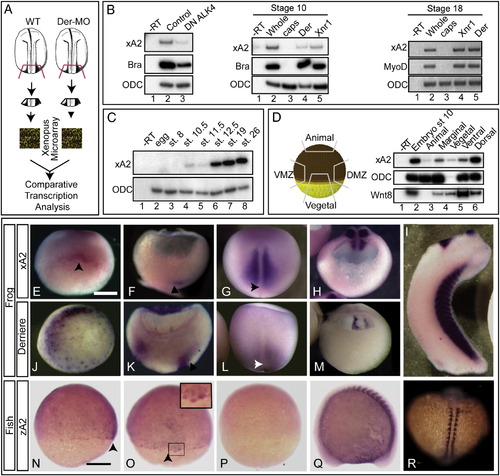
APOBEC2 is a target of TGFβ signaling coexpressed with derrière in Xenopus embryos. (A) Strategy for identification of genes regulated by derrière. Posterior dorsal fragments from wild-type and derrière -MO (Der-MO)-injected embryos were isolated at stage 18. (B) TGFβ signaling and xA2 expression. Overexpression of a dominant negative type 1 receptor (DN ALK4) reduces expression of marginal xA2 in stage 10 whole embryos (left panel). Overexpressed Xnr1 (30 pg) and derrière (100 pg) RNA induce xA2 expression in stage 10 (central panel) and 18 (right side panel) animal caps. RT-PCR for xA2, MyoD, and Brachyury as markers of mesoderm induction, and ODC as loading control. (C) Timing of xA2 expression. RT-PCR of embryos collected at the indicated developmental stages. (D) Spatial expression of xA2 in stage 10 embryos. RT-PCR of embryonic explants (VMZ: ventral marginal zone; DMZ: dorsal marginal zone). (E–M) Comparative expression of xA2 and derrière. In situ hybridization for xA2 (E–I) and derrière (J–M) expression. (E, J) Stage 10 vegetal–dorsal views, arrowhead indicates the forming dorsal lip; (F, K) stage 11 dorso-ventral sections (dorsal to the right). Arrowheads indicate recently involuted mesoderm; (G, L) dorsal views (anterior up). Arrowheads indicate the blastopore; (H, M) stage 16 transversal sections, posterior fragments (dorsal is up). (I) Stage 32 lateral view. Overlap between xA2 and derrière occurs at stage 10 (dorsal marginal), stage 11 (involuted mesoderm), and stage 16 (paraxial mesoderm). (N–R) Expression of zebrafish A2 (zA2). (N, O, P) Seventy-five percent epiboly, in (N) lateral view (dorsal to the right) and (O) dorsal views. The arrowheads in panels N and O indicate the shield. (Q–R) Fourteen somite stage embryo. The inset in panel O (twofold magnification) shows shield cells with nuclear stain. (Q) Lateral view, anterior to the left, (R) dorsal view (anterior up). (P) Embryo stained with the sense probe as negative control. The scale bars in panel E indicates 0.3 mm, and in panel N, 0.1 mm. EXPRESSION / LABELING:
|
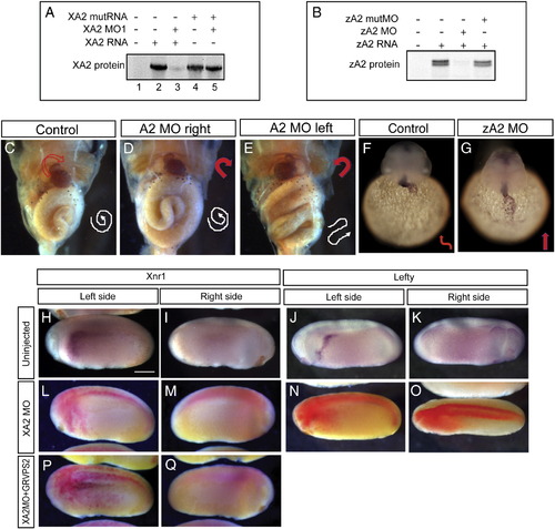
Effect of APOBEC2 protein depletion in Xenopus and zebrafish. (A, B) Inhibition of in vitro translation by xA2 MO (A) and zA2 MO (B). (C–E) Left-side depletion of xA2 protein randomizes the left–right axis in Xenopus. Embryos injected on the left side with 10 ng xA2 MO were stained for light meromyosin at stage 46. (C) Control embryo; (D) right-side MO injection normal embryo; (E) left-side MO injection, inverted heart and abnormally folded intestine. Arrows indicate the direction of the heart outflow tract and intestinal looping. (F–G) Depletion of zA2 protein prevents heart looping in zebrafish. In situ hybridization with cmlc2 antisense probe for heart muscle on 36–40 hpf embryos. Arrows indicate ventricular looping. (H–Q) xA2 depletion blocks the left-side nodal signal. In situ hybridization for Xnr1 (H, I, L, M, P, Q), and Lefty (J, K, N, O) in purple, and injected LacZ RNA as tracer (L–Q) in red. Wild-type expression of Xnr1 (H) and Lefty (J) in the left lateral plate mesoderm was inhibited by injection of xA2 MO in the left paraxial mesoderm (L, N). Left-side expression of Xnr1 was rescued by coinjection of GRVP16SMAD2Δ3 RNA (25 pg RNA, induced at stage 16; P). All views are lateral, except in panel O (dorsal), anterior to the left. Embryos are stage 23 (Xnr1) and stage 24 (Lefty). The scale bar in panel H represents 0.3 mm. |
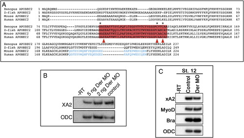
Sequence and expression of xA2. (A) Sequence of Xenopus, mouse, and zebrafish APOBEC2, and human APOBEC1. The active site ofAPOBEC1 is boxed. Active center residues, where H100, C130 and C134 coordinate Zn, are labeled with asterisks, and residues mutated in MutXA2 are indicated by red arrowheads. (B, C) xA2 expression is decreased in stage 18 posterior poles (B, 2 and 5 ng Der MO) and stage 12 dorsal halves (C, 5 ng Der MO) of Xenopus embryos depleted of Derriere protein. RT-PCR with ODC as loading control. |
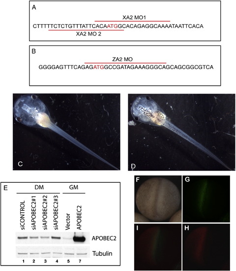
(A–E) Blocking reagents for Xenopus, zebrafish, and mouse apobec2. (A) Sequence of the two morpholino oligonucleotides for Xenopus A2 depletion. (B) Sequence of the morpholino oligonucleotide for zebrafish A2 depletion. (C, D) Unilateral PM injection of XA2 MO (10 ng) does not affect antero-posterior development. Dorsal views of control (C) and XA2 MO injection in left PM (D). (E) Effect of different siRNAs (1–3) against mouse A2 mRNA on expression of A2 protein levels in C2C12 cells. siA2.2 was chosen for the experiments in Fig. 5. DM: differentiation medium, GM: growth medium. Lanes 1–5: endogenous A2. Lane 7: overexpressed A2 (CMV-APOBEC2 transfection). Western blot for mouse A2 and tubulin. (F–I) Targeted injections for PM and LPM. Embryos injected at the 4 cell stage with fluorescent control MO (lateral subequatorial in the dorsal left blastomere, G) and Cherry-H2B (lateral subequatorial in the ventral left blastomere, H) were fixed at stage 20. Dorsal views, posterior is down. The same embryo in visible (F), green UV channel (G), red UV channel (H), and overlap of green and red (I). |
Reprinted from Developmental Biology, 350(1), Vonica, A., Rosa, A., Arduini, B., and Brivanlou, A.H., APOBEC2, a selective inhibitor of TGFβ signaling, regulates left–right axis specification during early embryogenesis, 13-23, Copyright (2011) with permission from Elsevier. Full text @ Dev. Biol.