
- Title
-
Characterization of pip5k3 fleck corneal dystrophy-linked gene in zebrafish
- Authors
- Boisset, G., Polok, B.K., and Schorderet, D.F.
- Source
- Full text @ Gene Expr. Patterns
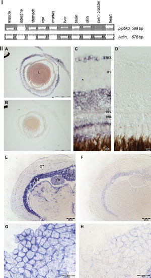
Expression profile of adult zebrafish pip5k3. (I) RT-PCR analysis of pip5k3 expression in different tissues of zebrafish. Pip5k3 was expressed in all tested tissues, but at low level in swim bladder. The house-keeping gene actin was used as control. The size of the amplified fragments is indicated on the right. (II, A–H) In situ hybridization using RNA probe specific for zebrafish pip5k3 on adult eye, brain and muscle cryosections. (A) Pip5k3 was expressed in the cornea and lens. (C) In the retina, pip5k3 staining was seen in GCL, INL and OLM. (E) In the brain, pip5k3 staining was mainly observed in PGZ, Val and TL. (G) Pip5k3 staining was perinuclear and located to the membranes of the cells in the muscle. (B, D, F and H) Control staining with sense probe. C, cornea; L, lens; GCL, ganglion cell layer; INL, inner nuclear layer; IPL, inner plexiform layer; LT, longitudinal torus; OLM, outer limiting membrane; ONL, outer nuclear layer; OPL, outer plexiform layer; OT, optic tectum; PGZ, periventricular gray zone of the optic tectum; Val, lateral division of valvula cerebelli. Scale bars: (A and B) 100 μm; (C and D and G and H) 10 μm; (E and F) 20 μm. EXPRESSION / LABELING:
|
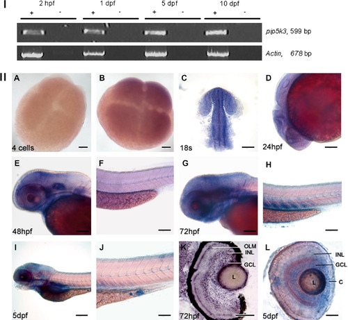
Embryonic expression of zebrafish pip5k3. (I) RT-PCR analysis of embryonic pip5k3 during development. Pip5k3 transcripts were detected at 2 hpf, 1 dpf, 5 dpf and 10 dpf. Absence of DNA contamination was controlled by a no reverse transcriptase experiment. The amplified fragments lengths are written on the right. (II) Whole mount in situ hybridization. (A) Control experiments using sense probe. (B, C, dorsal view and D, lateral view) pip5k3 was uniformly expressed at 4-cell stage, 18 somites and 24 hpf. (E, G and I) Expression of pip5k3 in the head region at 48 hpf, 72 hpf and 5 dpf. Note that the staining in the gut region was not specific compared to the sense probe (I and data not shown). (F, H and J) View of the tail of 48 hpf, 72 hpf and 5 dpf embryos. No staining was detected at 48 hpf in contrast to 72 hpf and 5 dpf where a partial expression of pip5k3 was found in somites. Note that the blue spot localized to the proctodeum at 5 dpf was an artefact (J). (K) In situ on 12 μm cryosectionned eye of 3 dpf-old larva. Staining was mainly observed in OLM, INL, GCL and L. (L) Eye of 5 dpf-old larva showing expression of pip5k3 in INL, GCL, L and C. OLM, outer limiting membrane; INL, inner nuclear layer; GCL, ganglion cell layer; L, lens; C, cornea. Scale bars: (A and B) 20 μm; (C–H and J) 100 μm; (I) 200 μm; (K and L) 50 μm. |
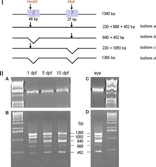
Spliced variants analysis during development. (I) Experimental procedure. Restriction site for HindIII and MslI in exon 2 and 7 are shown (arrows). Length of each fragments found after digestion for all isoforms are shown on the right. (II, A and C) Larval cDNA and adult eye cDNA before digestion. (II, B) Digested larval cDNA at stage 1 dpf, 3 dpf and 5 dpf showed a steady state expression of isoform a (668 bp) and isoform d (1265 bp). Isoform b (840 bp) is upregulated and Isoform c (1093 bp) is downregulated. (II, D) Isoforms a, b, c and d are present in adult eye, isoform b and d at high level. |
Reprinted from Gene expression patterns : GEP, 8(6), Boisset, G., Polok, B.K., and Schorderet, D.F., Characterization of pip5k3 fleck corneal dystrophy-linked gene in zebrafish, 404-410, Copyright (2008) with permission from Elsevier. Full text @ Gene Expr. Patterns