
- Title
-
Expression of cyp26b1 during zebrafish early development
- Authors
- Zhao, Q., Dobbs-McAuliffe, B., and Linney, E.
- Source
- Full text @ Gene Expr. Patterns
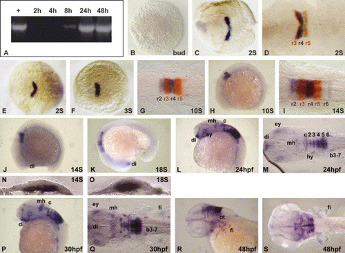
The distribution of cyp26b1 message during early development of zebrafish. (A) RT-PCR results showing the presence of cyp26b1 message within 48 hpf. Lane 1 is a positive control using the cDNA as a template (+). No expression was detected at either 2 or 4 h (hours), but weak expression of cyp26b1 was detected at 8 h (75% epiboly). (B-S). Whole mount in situ hybridization showing expression of cyp26b1 message in zebrafish during early development (cyp26b1—blue, krox20—red). (B—K, M, Q—S) anterior left. (L, P) anterior top. (B) lateral view, 10 hpf (bud stage), no expression was detected. (C) dorsal view, 10.5 hpf (2-somites (S)), cyp26b1 expression in two stripes lateral to the midline. (D) dorsal view of 10.5 hpf embryo (flat-mount), cyp26b1 is expressed in r4 and has overlapping expression with krox20 in r3. (E) dorsal view, 10.7 hpf (2 S). (F) dorsal view, 11 hpf (3S). (G) flatmount, dorsal view of the 14 hpf embryo (10S). Expression of cyp26b1 covers r2, r3 and r4. (H) lateral view, 14 hpf (10S). (I) dorsal view (flatmount), hindbrain of a 16 hpf (14S) embryo, expression of cyp26b1 extends from r2 and r4, boundaries of r5 and r6, centering at r4. (J) lateral view, 16hpf (14S) embryo, weak cyp26b1 expression is also seen in the diencephalon. (K) lateral view of 18 hpf embryo, expression of cyp26b1 covers r2 through r6 and expression remains in the diencephalon. (L) lateral view of 24 hpf embryo. (M) dorsal view of 24 hpf (flat-mount) embryo, expression of cyp26b1 is present in diencephalon, eyes, midbrain hindbrain boundary, cerebellum, boundaries of r2 and r3, rhombere 4, 5 and 6, the hyoid arch and branchial arches 3–7. (N) dorsal top, cross section of the hindbrain of a 14S embryo. Cyp26b1 expression is limited to the ventral portion of the hindbrain. (O) dorsal top, cross section of the hindbrain of an 18S embryo. Again, expression is in the ventral portion of the hindbrain. (P) lateral view of a 30 hpf embryo. (Q) dorsal view of a 30 hpf embryo (flat-mount), expression of cyp26b1 is present in diencephalon, eyes, midbrain hindbrain boundary, cerebellum, boundaries of r2, r3 and r4, rhombere 5 and 6, the hyoid arch, branchial arch primordia 3–7, and the pectoral fins. (R) lateral view of 48 hpf embryo. (S) dorsal view of 48 hpf embryo, expression of cyp26b1 is present in the diencephalon, eyes, midbrain hindbrain boundary, cerebellum, hindbrain r2–r6, the hyoid arch, the branchial arches, and pectoral fins. b: brachial arch primordia, c: cerebellum, di: diencephalon, ey: eye, fi: pectoral fin, h: hyoid arch, mh: midbrain hindbrain boundary, ot: otic vesicle, r: rhombomere. |

Unillustrated author statements EXPRESSION / LABELING:
|
Reprinted from Gene expression patterns : GEP, 5(3), Zhao, Q., Dobbs-McAuliffe, B., and Linney, E., Expression of cyp26b1 during zebrafish early development, 363-369, Copyright (2005) with permission from Elsevier. Full text @ Gene Expr. Patterns