
Fig. 3
- ID
- ZDB-IMAGE-260921-23
- Publication
- Reitz et al., 2026 - Molecular characterization of FAM222B as a novel disease gene for dominant cardiovascular laterality defects
- All Figures
- Figures for Reitz et al., 2026
|
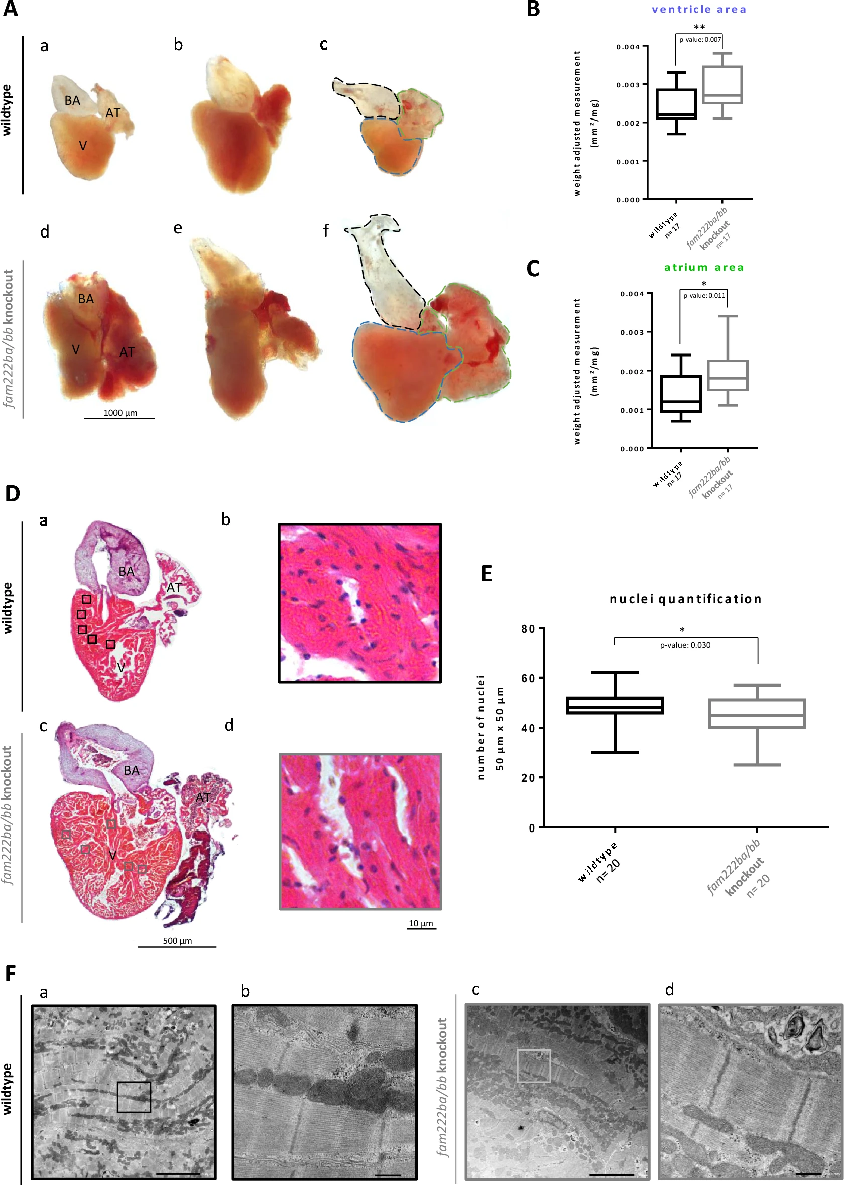
Fig. 3 Examination of adult zf hearts, morphology and histology. (A) Detailed examination and comparison of dissected adult wt and fam222ba/bb dd-KO zf ‘ hearts (N = 4, n = 17 in each group, except for BA analysis in dd-KO zf group: N = 4, n = 16, in total): (a-c) show representative examples of normally developed wt zf hearts, (d-f) demonstrate various representative altered fam222ba/bb dd-KO zf hearts. (d) presents a hypertrophic atrium, (e) shows a slim ventricle and (f) reveals a hypertrophic BA. Scale bar: 1000 µm. Dashed lines symbolize the way of measurement exemplary in c&f for the zf ‘ ventricle in blue, the atrium in green and the BA in black. Abbreviations: AT = atrium, BA = bulbus arteriosus, V = ventricle. No significant difference between BA area of wt and fam222ba/bb dd-KO zf. Median (mm2/ mg): wt: 0.0010, dd-KO: 0.0011. Unpaired two-tailed t-test: p-value: 0.366, ns = not significant. (B) Box plot graph comparing wt and fam222ba/bb dd-KO zf ‘ ventricle area. Median (mm2/ mg): wt: 0.0022, dd-KO: 0.0027. Unpaired two-tailed t-test: p-value: 0.007, ** = very significant. (C) Box plot graph comparing wt and fam222ba/bb dd-KO zf ‘ atrium area. Median (mm2/ mg): wt: 0.0012, dd-KO: 0.0018. Unpaired two-tailed t-test: p-value: 0.011, * = significant. (D) HE-Staining of adult wt (a,b) and dd-KO (c,d) zf hearts. Each box in the overview (a,c) corresponds to a 50 µm × 50 µm area used for quantification of nuclei. Both areas enclosed by thicker (bold) boxes are presented examples at higher resolution in the corresponding magnified panel (b,d). Scale bar: overview: 500 µm, zoom: 10 µm. Abbreviations: AT = atrium, BA = bulbus arteriosus, V = ventricle. (E) Box plot graph comparing the number of nuclei in each selected wt and fam222ba/bb dd-KO heart in five 50 µm × 50 µm areas of ventricular myocardium (N = 4, n = 20). Median: wt: 48, dd-KO: 45. Unpaired two-tailed t-test: p-value: 0.030, * = significant. (F) TEM images showing ventricular myocardium of adult wt (a,b) and fam222ba/bb dd-KO (c,d) zf. Overview (a,c) and higher magnification (b,d) of sarcomeres. Box in overview indicates zoomed section. There was no apparent difference neither in the overall tissue assembly nor in the detailed composition of sarcomeres. Scale bar: overview: 5 µm, zoom: 500 nm.