
Fig. 7
- ID
- ZDB-IMAGE-260723-44
- Publication
- Kaveh et al., 2026 - Flow-mediated endothelial remodeling and inflammation drive developmental vascular susceptibility in ldlr loss of function
- All Figures
- Figures for Kaveh et al., 2026
|
Fig. 7
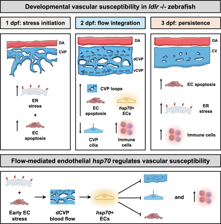
Schematic model of developmental vascular susceptibility in ldlr-/- zebrafish.
At 1 dpf (stress initiation), ldlr-/- embryos exhibit elevated endoplasmic reticulum (ER) stress signaling accompanied by increased endothelial apoptosis in trunk vessels (top, left). At 2 dpf (flow integration), abnormal dorsal caudal venous plexus (CVP) angiogenic remodeling is observed, spatially associated with the emergence of flow-dependent hsp70+ endothelial cells (ECs). This stage is accompanied by sustained vascular apoptosis, increased CVP loop formation, reduced CVP ciliogenesis and enhanced expansion of innate immune cells within the caudal hematopoietic tissue. Dashed arrows indicate CVP blood flow direction and relative magnitude (top, middle). At 3 dpf (persistence), CVP remodeling remains partially disrupted, while vascular apoptosis, ER stress signaling and immune cell expansion persist (top, right). This model proposes that early endothelial stress converges with physiological blood flow through a structurally altered CVP to drive hsp70 upregulation and modulate vascular susceptibility. The emergence of hsp70 + ECs promotes endothelial survival while inhibiting endothelial ciliogenesis and angiogenic remodeling (bottom). Partly created in BioRender. Kaveh, A. (2026) https://BioRender.com/xft5lm8. DA, dorsal aorta; dCVP, dorsal caudal venous plexus; vCVP, ventral caudal venous plexus; CV, caudal vein.