
Fig. 2
- ID
- ZDB-IMAGE-260607-2
- Publication
- Perey-Simon et al., 2025 - dUTPase is essential in zebrafish development and possesses several single-nucleotide variants with pronounced structural and functional consequences
- All Figures
- Figures for Perey-Simon et al., 2025
|
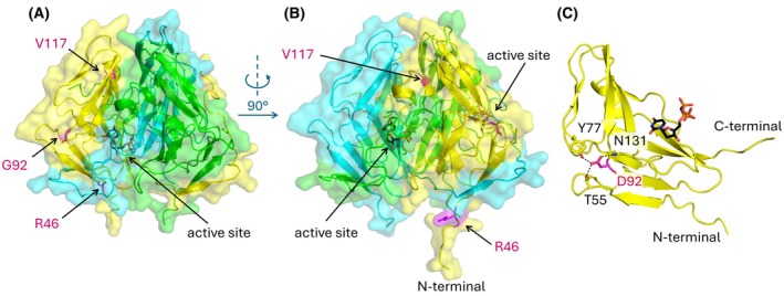
Fig. 2
Spatial arrangement of Danio rerio dUTPase mutations relative to the surface and the active site of the protein. (A, B) Surface of DrDUT‐RGV (Alphafold 3 model) is shown (partially transparent) from two different viewpoints (rotated around the y axis by 90 degrees) to allow inspection of all three positions. Protein chains are colored yellow, green and cyan, positions of the variable residues in case of Chain A (yellow) highlighted with magenta. The position of the substrate analogue (shown as sticks with atomic coloring carbon : black, oxygen : red, nitrogen : blue, phosphorus : orange) at the active site is modeled based on superimposition of DrDUT‐RGV with the human dUTPase (PDB ID: