
Fig. 8
- ID
- ZDB-IMAGE-260528-14
- Genes
- Publication
- Ghosh et al., 2025 - EIPR1 variants cause a neurodevelopmental disorder with endolysosomal and dense core vesicle defects
- All Figures
- Figures for Ghosh et al., 2025
|
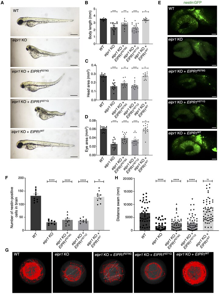
Fig. 8 Human wild-type (WT) but not EIPR1 variant mRNAs rescue neural progenitor cells and motor function in eipr1 knockout (KO) zebrafish. (A) Morphology of WT, eipr1 KO, eipr1 KO + EIPR1R279G, eipr1 KO + EIPR1H271Q and eipr1 KO + EIPR1WT larvae at 3 days post-fertilization (3 dpf). Scale bar = 1 mm. (B–D) Quantification of body length (B), head area (C) and eye area (D) from larvae such as those shown in A (N = 3, n = 14–19 for body length, n = 9–14 for head area, n = 11–16 for eye area). (E) Representative images of neural progenitors (nestin:GFP) in WT, eipr1 KO, eipr1 KO + EIPR1R279G, eipr1 KO + EIPR1H271Q and eipr1 KO + EIPR1WT brain at 1 dpf. Scale bar = 40 µm. (F) Quantitative analysis demonstrated a significant decrease in the neural progenitors (nestin:GFP) in eipr1 KO, eipr1 KO + EIPR1R279G (932) and eipr1 KO + EIPR1H271Q zebrafish compared with WT zebrafish, but no significant change was observed in eipr1 KO + EIPR1WT zebrafish at 1 dpf, n = 8–10. (G) Representative swimming tracks of WT, eipr1 KO, eipr1 KO + EIPR1R279G, eipr1 KO + EIPR1H271Q and eipr1 KO + EIPR1WT zebrafish at 5 dpf. (H) Quantification of swimming distance in WT, eipr1 KO, eipr1 KO + EIPR1R279G, eipr1 KO + EIPR1H271Q and eipr1 KO + EIPR1WT zebrafish at 5 dpf. All data are presented as the mean ± SEM. Statistical significance was calculated by Student’s t-test, or one-way ANOVA followed by Dunnett’s T3 multiple comparisons test. **P < 0.01, ***P < 0.001, ****P < 0.0001. GFP = green fluorescent protein.