IMAGE

Figure 2.

ID
ZDB-IMAGE-260423-46
Publication
Wei et al., 2026 - A Novel CRYBB2 Splicing Mutation Is Associated With Lens Extracellular Matrix Remodeling and Vascular Alterations in Congenital Cataract
All Figures
Figures for Wei et al., 2026
Image
Figure Caption

Figure 2.

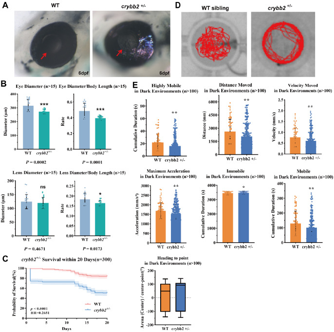
Characterization of WT and crybb2+/− zebrafish larvae. (A) Stereomicroscopic imaging of WT and crybb2+/− zebrafish eyes at 6 dpf. Images were captured using a Leica M205 FA stereomicroscope. (B) Measurements of eye and lens diameters and their ratios to body length in zebrafish larvae at 6 dpf. Quantification was performed on 10 larvae per group. Images were processed and analyzed using ImageJ software. (C) Survival analysis of WT and crybb2+/− larvae from 1 to 20 dpf. Survival curves were plotted and analyzed using GraphPad Prism 8 with the Kaplan-Meier method. (D) Representative locomotor trajectories of zebrafish larvae at 7 dpf. (E) Quantitative analysis of locomotor activity at 7 dpf, including traveled distance, swimming speed, maximum acceleration, activity states, and resting time. Behavioral tracking was conducted using EthoVision XT software (Noldus Information Technology, Leesburg, VA, USA).

Figure Data
Acknowledgments
This image is the copyrighted work of the attributed author or publisher, and ZFIN has permission only to display this image to its users. Additional permissions should be obtained from the applicable author or publisher of the image. Full text @ Invest. Ophthalmol. Vis. Sci.