IMAGE

FIGURE 5

ID
ZDB-IMAGE-260129-40
Source
Figures for Han et al., 2026
Image
Figure Caption

FIGURE 5

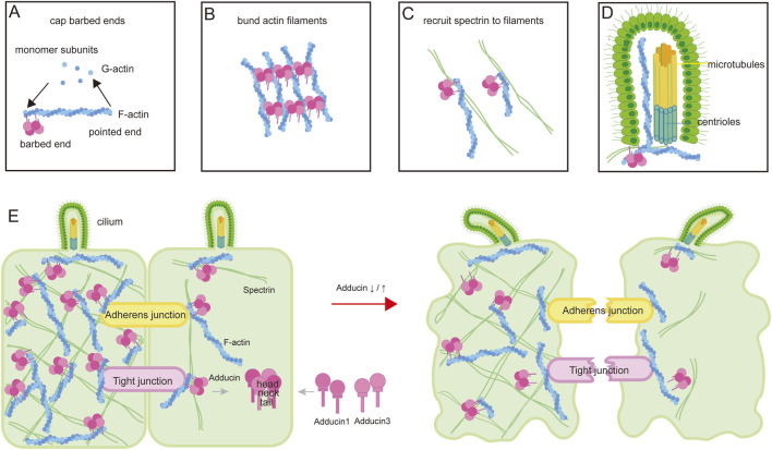
A schematic diagram illustrating the function of ADD3 and the molecular mechanisms by which ADD3 is involved in BA pathogenesis. (A) The actin cytoskeleton is composed of actin filaments polymerized from monomeric G-actin. Adducin, typically a heterodimer of ADD1 and ADD3, caps the barbed ends of actin filaments to inhibit further polymerization. (B) Adducins facilitate bundling of actin filaments. (C) Adducins recruit and crosslink spectrin at the filament termini, organizing the spectrin-actin network. (D) The cytoplasmic actin-adducin cytoskeleton regulates centrosome migration and docking to the apical plasma membrane, facilitates vesicle transport to the centriole, and mediates the entry of signal receptors into the ciliary membrane. Additionally, F-actin is a component of the axonemal cytoskeleton, positioned between alpha-tubulin singlets and located between microtubules and the ciliary membrane. (E) Adducins play a crucial role in maintaining cell shape, stabilizing junction complexes, and regulating ciliogenesis and ciliary function (left panel). Dysregulation of Adducin alters cell shape, impairs the stability of tight junctions and cell barrier integrity, disrupts cilia structure and function, and thereby contributes to BA pathogenesis (right panel).

Acknowledgments
This image is the copyrighted work of the attributed author or publisher, and ZFIN has permission only to display this image to its users. Additional permissions should be obtained from the applicable author or publisher of the image. Full text @ Front Genet