
|
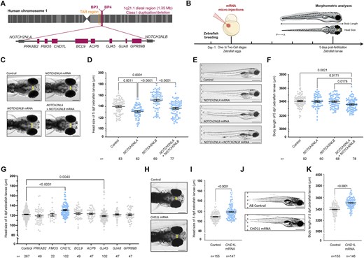
Fig. 1 In vivo overexpression of the genes from the 1q21.1 distal region in zebrafish. (A) Schematic representation of the human 1q21.1 BP3–BP4 distal region showing eight genes comprised in the region and NOTCH2-paralogs NOTCH2NLA and NOTCH2NLB located at the breakpoints. (B) Schematic representation of the experimental procedure including micro-injection of mRNA in one- to two-cell stage zebrafish embryos and morphometric analyses at 5 dpf for the head size and the body length of zebrafish larvae. (C) Dorsal views of the head of control larvae and larvae injected with NOTCH2NLA mRNA (50 pg), NOTCH2NLB mRNA (50 pg) or NOTCH2NLA+ NOTCH2NLB mRNA (50 + 50 pg) at 5 dpf. Double-ended arrows indicate distance between the eyes. (D) Dot plot showing the distance between the eyes (head size) of control larvae and larvae injected with NOTCH2NLA mRNA (50 pg), NOTCH2NLB mRNA (50 pg), or NOTCH2NLA+ NOTCH2NLB mRNA (50 + 50 pg) at 5 dpf. Data shown as mean ± SEM of a least triplicate batches; Kruskal–Wallis test. (E) Lateral view of control larvae and larvae injected with NOTCH2NLA mRNA (50 pg), NOTCH2NLB mRNA (50 pg) or NOTCH2NLA+ NOTCH2NLB mRNA (50 + 50 pg) at 5 dpf. (F) Dot plot showing the body length measurements of control larvae and larvae injected with NOTCH2NLA mRNA (50 pg), NOTCH2NLB mRNA (50 pg), or NOTCH2NLA+ NOTCH2NLB mRNA (50 + 50 pg) at 5 dpf. Data shown as mean ± SEM of at least triplicate batches per condition; ordinary one-way analysis of variance (ANOVA) test. (G) Dot plot showing the distance between the eyes (head size) of 5 dpf control larvae and larvae injected with each of the eight human genes from the 1q21.1 distal region. Data shown as mean ± SEM of a least triplicate batches per gene; Kruskal–Wallis test. (H) Dorsal views of control and larvae injected with CHD1L mRNA (100 pg) at 5 dpf. Double-ended arrows indicate distance between the eyes. (I) Dot plot showing the distance between the eyes (head size) of control and CHD1L mRNA-injected larvae (100 pg) at 5 dpf. Data shown as mean ± SEM of a least triplicate batches; Mann–Whitney test. (J) Lateral views of control and CHD1L mRNA-injected larvae (100 pg) at 5 dpf. (K) Dot plot showing the body length measurements of control or injected larvae with CHD1L mRNA (100 pg) at 5 dpf. Data shown as mean ± SEM of triplicate batches; Mann–Whitney test. To simplify the visualization of the data, the nonsignificant P-values are not shown on the graphs. Scale bars: 200 μm [panels (C) and (H)] and 500 μm [panels (E) and (J)].