
|
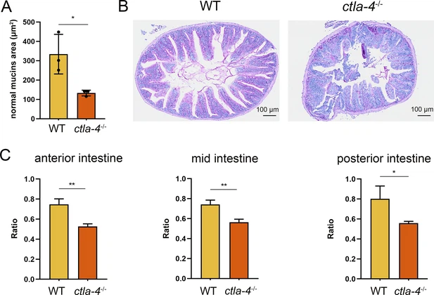
Fig. 2 - Supplemental 1 Histopathological analysis of intestines. (A) Quantitative analysis of the mucins area stained by Alcian Blue and Periodic Acid-Schiff (AB-PAS) from WT and ctla-4-/- zebrafish (n=3) by ImageJ software (version 1.8.0). (B) Periodic Acid-Schiff (PAS) staining was used to analyze the mucin components in anterior intestine from wild-type (WT) and ctla-4-/- zebrafish (n=5). (C) The ratio of intestinal villi length to intestinal ring radius was measured in the anterior, mid, and posterior intestines of WT and ctla-4-/- zebrafish (n=6). Statistical significance was assessed through an unpaired Student’s t-test (*p < 0.05; **p < 0.01).