
Image
|
Figure Caption
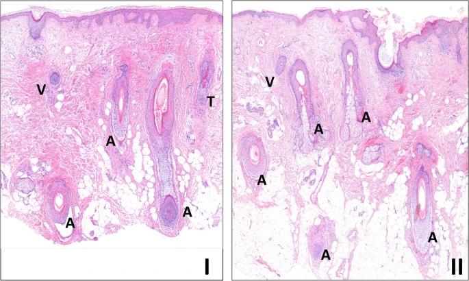
Fig. 3 The effects in the skin biopsies. Histology of skin biopsies before (I) and after (II) treatment, taken from the scalp at the hairline of case 2 (H&E stained, 100x), reveals essentially identical features, as assessed by the pathologist at our center, who has experience in evaluating such skin biopsies, in collaboration with the involved dermatologists. The hair follicles in both samples are predominantly in the anagen phase (A). In the biopsy before treatment (I), one hair follicle in the telogen phase (T) and one vellus hair follicle (V) are also visible.
Acknowledgments
This image is the copyrighted work of the attributed author or publisher, and
ZFIN has permission only to display this image to its users.
Additional permissions should be obtained from the applicable author or publisher of the image.
Full text @ Sci. Rep.