
|
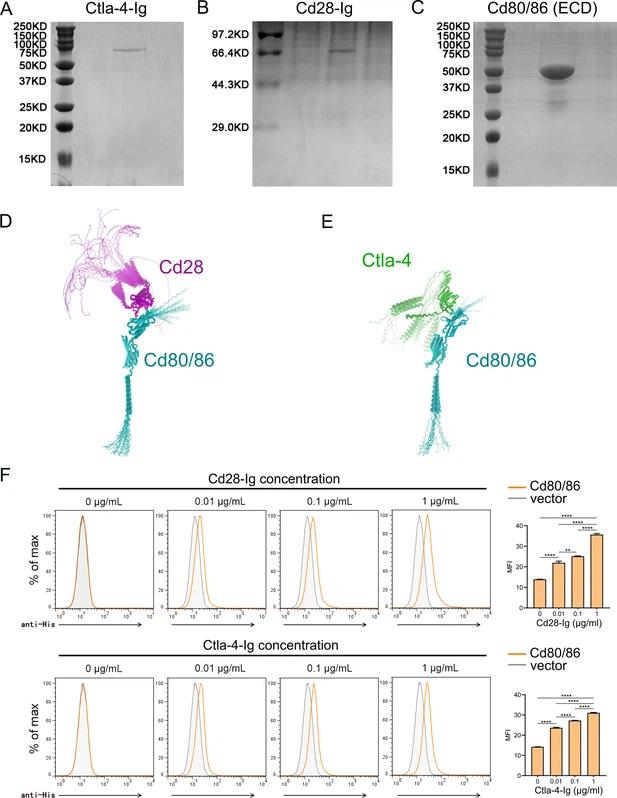
Fig. 7 - Supplemental 1 Preparation of recombinant proteins and examination of their molecular interactions. (A–C) SDS-PAGE detection of the purified recombinant soluble Ctla-4-Ig (sCtla-4) (A) and sCd28-Ig (B) proteins and the Cd80/86 extracellular domain (ECD) (C) with Coomassie brilliant blue staining. (D–E) The predicted molecular interactions between Cd80/86 and Cd28 (D), as well as Cd80/86 and Cytotoxic T lymphocyte antigen-4 (Ctla-4) (E), as modeled by AlphaFold2. The structures are represented in a cartoon style, with Cd80/86, Cd28, and Ctla-4 colored cyan, green, and magenta, respectively. A total of 25 models were predicted for each complex and aligned with Cd80/86. (F) Flow cytometry analysis of the interactions between Cd80/86 and Cd28 (top), and Cd80/86 and Ctla-4 (bottom). Cd80/86 was expressed on HEK293T cells and incubated with varying concentrations of fluorescently labeled sCd28-Ig or sCtla-4-Ig. Fluorescence intensity was detected by flow cytometry to determine molecular interactions. Data are presented as mean ± SD, derived from three independent experiments. Statistical significance was evaluated using an unpaired Student’s t-test (**p < 0.01; ***p < 0.001).