
|
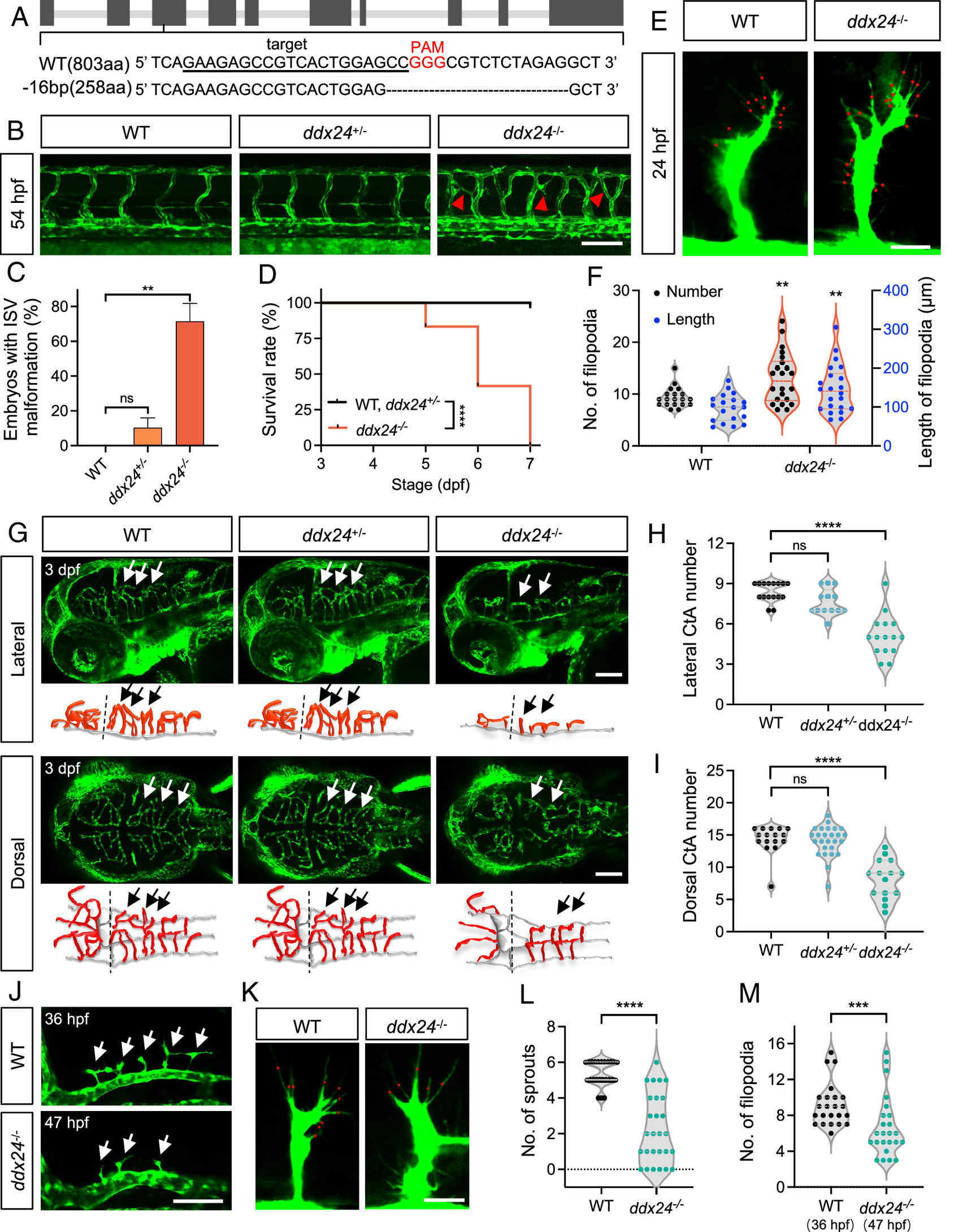
Fig. 2 Loss of Ddx24 promotes endothelial cell branching in the trunk and impairs sprouting in the brain of zebrafish. (A) CRISPR/Cas9 target site in ddx24 locus with 16-bp deletion (PAM in red). (B) Representative confocal images of trunk vasculature in WT, ddx24+/-, and ddx24−/− embryos at 54 hpf. Red arrowheads indicate ectopic ISV branches. (C) Quantification of the embryo with ectopic ISV branches. n = 29, 40, 23 embryos. (D) Kaplan–Meier survival curves of WT, ddx24+/-, and ddx24−/− zebrafish. n = 18, 36, 12 embryos. (E) Representative high-magnification confocal images of ISV sprouts in WT and ddx24−/− embryos at 24 hpf. The red dots point to the tip cell filopodia. (F) Quantification of filopodia number and length. n = 19 for WT, n = 22 for ddx24−/− embryos. (G) Representative confocal images and schematic diagram of hindbrain vasculature in WT, ddx24+/-, and ddx24−/− embryos at 3 dpf. Arrows indicate CtAs. (H and I), Quantification of CtAs. Lateral view number of CtAs (H, n = 16, 13, 15 embryos) and dorsal view number of CtAs (I, n = 16, 26, 15 embryos) were determined. (J) Representative confocal images of hindbrain vasculature in WT (36 hpf) and ddx24−/− (47 hpf) embryos. Arrows indicate CtAs. (K) Representative high-magnification confocal images of CtA sprouts in WT (36 hpf) and ddx24−/− (47 hpf) embryos. The red dots point to the tip cell filopodia. (L and M), Quantification of CtA sprouts (L, n = 24, 27 embryos) and filopodia per CtA (M, n = 23, 24 embryos). [Scale bars, 100 μm in (B, G, and J); 20 μm in (E and K).] Data are represented as mean ± SD. ns, not significant, **P < 0.01, ***P < 0.001 and ****P < 0.0001, as assessed by one-way ANOVA (C), log-rank (Mantel–Cox) test (D) parametric two-tailed Student’s t test (F), nonparametric Kruskal–Wallis test (H and I) and nonparametric Mann–Whitney test (L and M).