IMAGE

Figure 9

ID
ZDB-IMAGE-250530-38
Source
Figures for Zheng et al., 2025
Image
Figure Caption

Figure 9

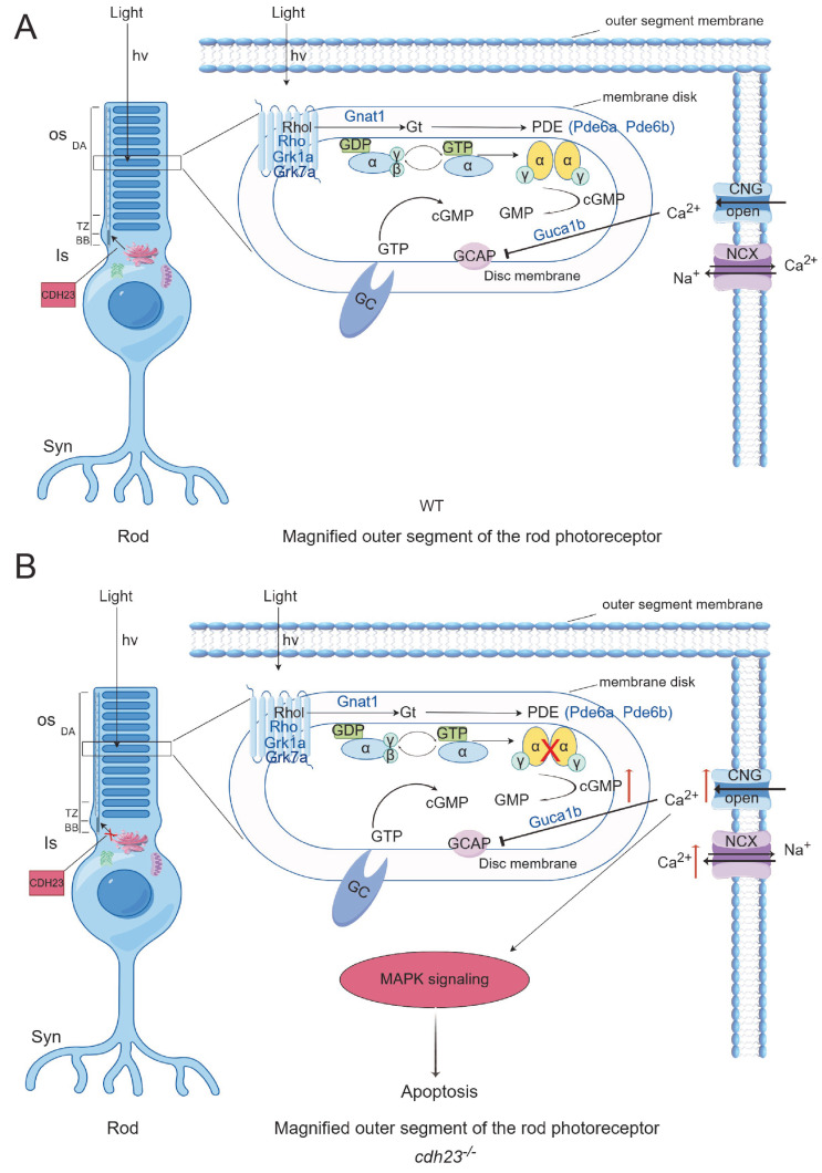
Schematic diagram of cdh23 regulating rod cell calcium ion transport (image created by Figdraw). (A) In WT rod photoreceptor cells, genes such as Rho, Rhol, Grk1, Grk7a, Gnat1, Pde6a, Pde6b, and Guca1b play essential roles in phototransduction. Additionally, Atp2b1b is responsible for transport of calcium ions (Ca2+). Rhodopsin is synthesized in endoplasmic reticulum of inner segment (IS) and transported to outer segment (OS) through vesicular transport from Golgi apparatus, with IS and OS being connected by connecting cilium. Purine metabolism generates sufficient ATP to properly position rhodopsin in outer segment. Upon light stimulation, rhodopsin undergoes conformational change, activating Gt transducin protein, which subsequently activates phosphodiesterase (PDE). PDE hydrolyzes cGMP, leading to decrease in its concentration, which causes closure of CNG channels. This reduces inward flow of Ca2+ and release of neurotransmitter glutamate, completing phototransduction process. (B) In cdh23−/− embryos, disrupted purine metabolism leads to insufficient ATP production, impairing vesicle transport from Golgi apparatus and movement of cilium. As a result, rhodopsin fails to localize properly to outer segment, preventing cGMP hydrolysis by PDE. Increased cGMP concentration activates CNG channels, causing continuous inward flux of Ca2+, which activates Ca2+-dependent MAPK signaling pathway and triggers apoptosis in rod photoreceptor cells.

Acknowledgments
This image is the copyrighted work of the attributed author or publisher, and ZFIN has permission only to display this image to its users. Additional permissions should be obtained from the applicable author or publisher of the image. Full text @ Int. J. Mol. Sci.