
Image
|
Figure Caption
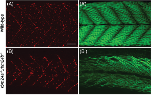
Fig. 10 Formation of neuromuscular junctions in rbm24a and rbm24b mutants. (A, B) Alpha-bungarotoxin staining. (A′, B′) Phalloidin staining. Lateral view of wild-type and rbm24a−/−;rbm24b−/− mutant embryos at 4 dpf. Notice that myofibers appear dispersed and disorganized in rbm24a and rbm24b mutant embryos, whereas neuromuscular jonctions are present and are arborized in a smilar pattern as those vizualized in the wild-type. Scale bar: 50 μm.
Figure Data
Acknowledgments
This image is the copyrighted work of the attributed author or publisher, and
ZFIN has permission only to display this image to its users.
Additional permissions should be obtained from the applicable author or publisher of the image.
Full text @ Dev. Dyn.