
|
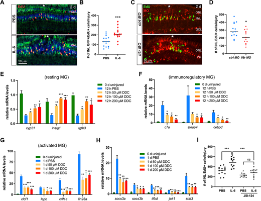
Fig. 4 IL-6 regulates MG reprogramming and proliferation via Stat3 signaling. (A) EdU immunofluorescence showing the cell proliferation in retinas of the Tg(gfap:GFP) fish at 2 dpi. (B) Quantification of the number of INL GFP+ EdU+ cells per injury of A. (C) EdU immunofluorescence showing MG proliferation in the INL at 2 dpi. Retinas were electroporated with 1 mM of control or il6r MO at the time of the stab injury. (D) Quantification of the number of INL EdU+ cells per injury of C. (E, F, G, H) The qPCR analysis of the expression levels of MG subtype markers and Jak1-Stat3 signaling components in retinas treated with PBS or indicated doses of DDC. Day 0 served as the uninjured control. The statistical analyses were between DDC groups and PBS control. (I) JSI-124 treatment abolished the promoting effect of IL-6 on MG proliferation at 4 dpi. White *, site of the stab injury. *, P < 0.05; **, P < 0.01; ***, P <0.001; DDC, Di-O-demethylcurcumin; GCL, ganglion cell layer; INL, inner nuclear layer; ns, non-significant; ONL, outer nuclear layer.