
|
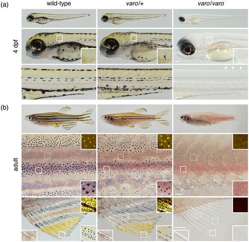
Fig. 1 Phenotypes of varo mutant. (a) Early larvae had irregular patterns of melanophores if heterozygous and lacked melanophores and xanthophores if homozygous. Insets show normal yellow color from xanthophores, posterior to otocyst, ectopic and hypomelanized melanophores, with yellow color of xanthophores in heterozygote and absence of melanophores and xanthophore color in homozygote. Arrowheads, persisting iridophores in normal locations in the homozygote, as a sheet of cells above the swim bladder and as individual cells along dorsal midline. Counts of dorsal iridophores did not differ among wild-type, heterozygous or homozygous siblings (F2,36 = 1.07, p = 0.4). (b) Adult heterozygotes had irregular stripes with melanophores that contained less melanin than the wild type, whereas homozygotes lacked stripes as well as melanophores and xanthophores. Details of flanks and fins are shown after treating with epinephrine to contract pigment granules towards cell centers. Flank insets: Details of areas outlined by dashed squares show pseudocolored xanthophores in bright yellow–orange on a dark background (Figure S1a); details of areas outlined by solid squares illustrate melanophores, iridophores (iridescent or purplish grey) or both. Fin insets: Dashed squares, pseudocolored xanthophores; solid squares, melanophores and xanthophores; dashed rectangles, iridophores (iridescent or purplish, examples circled), and xantholeucophores (orange–brown). Scale bars: A, 100 μm; b, 200 μm flank, 500 μm fin.