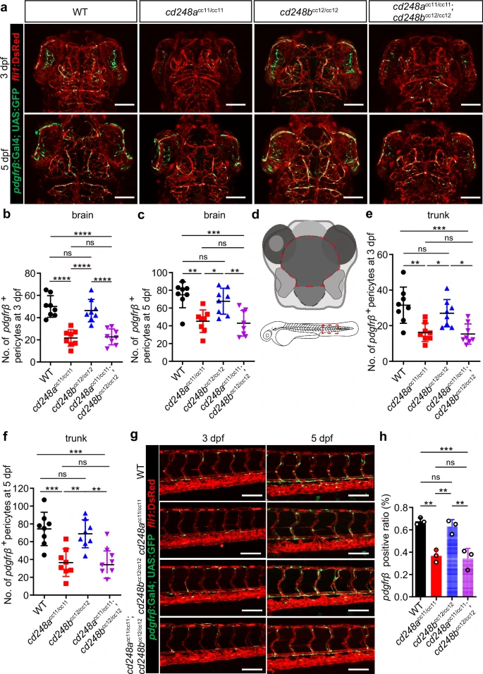
Fig. 4 Deficit of pericytes in cd248a mutant zebrafish.Confocal micrograph (a) and quantification of pdgfrβ+ brain pericytes at 3 dpf (b) and 5 dpf (c) in wild-type (WT) and mutant zebrafish (n = 8 embryos or larvae). Scale bars = 100 μm. d Schematic diagram of the counting region, as circled by the red dashed lines. Confocal micrograph (g) and quantification of pdgfrβ+ pericytes in the trunk region at 3 dpf (e) and 5 dpf (f) in wild-type (WT) and mutant zebrafish (n = 8 embryos or larvae). Scale bars = 100 μm. h FACS analysis of the proportion of pdgfrβ+ cells in wild-type and mutant fishes (n = 3 biologically independent experiments). Data were analyzed by One-way ANOVA and presented as mean ± SD. *, p < 0.05. **, p < 0.01. ***, p < 0.001.****, p < 0.0001. ns: no significance.
                
                    
                        Image


                        Figure Caption


                        Figure Data


                        Acknowledgments


This image is the copyrighted work of the attributed author or publisher, and
ZFIN has permission only to display this image to its users.
Additional permissions should be obtained from the applicable author or publisher of the image.
      Full text @ Commun Biol