
|
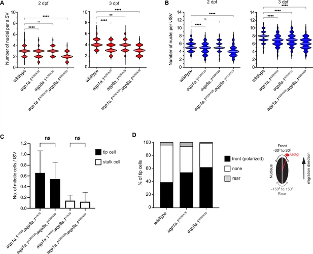
Fig. 4 - Supplemental 1 Reduction in endothelial cell (EC) number per ISV in aqp1a.1rk28/rk28;aqp8a.1rk29/rk29 embryos. (A) Quantification of EC number in arterial ISVs (aISVs) of wildtype and aquaporin mutant embryos at 2 dpf (wildtype: n = 273 aISVs from 31 embryos; aqp1a.1rk28/rk28: n = 189 aISVs from 21 embryos; aqp8a.1rk29/rk29: n = 84 aISVs from 10 embryos; aqp1a.1rk28/rk28;aqp8a.1rk29/rk29: n = 65 aISVs from 13 embryos) and 3 dpf (wildtype: n = 98 aISVs from 10 embryos; aqp1a.1rk28/rk28: n = 86 aISVs from 20 embryos; aqp8a.1rk29/rk29: n = 175 aISVs from 22 embryos; aqp1a.1rk28/rk28;aqp8a.1rk29/rk29: n = 102 aISVs from 22 embryos). Data collected from three (wildtype) and two (aquaporin mutants) independent experiments. (B) Quantification of EC number in venous ISVs (vISVs) of wildtype and aquaporin mutant embryos at 2 dpf (wildtype: n = 266 vISVs from 31 embryos; aqp1a.1rk28/rk28: n = 143 vISVs from 21 embryos; aqp8a.1rk29/rk29: n = 121 vISVs from 10 embryos; aqp1a.1rk28/rk28;aqp8a.1rk29/rk29: n = 95 vISVs from 13 embryos) and 3 dpf (wildtype: n = 121 vISVs from 10 embryos; aqp1a.1rk28/rk28: n = 143 vISVs from 20 embryos; aqp8a.1rk29/rk29: n = 280 vISVs from 22 embryos; aqp1a.1rk28/rk28;aqp8a.1rk29/rk29: n = 160 vISVs from 22 embryos). Data collected from three (wildtype) and two (aquaporin mutants) independent experiments. (C) Quantification of number of mitotic events per ISV in aqp1a.1+/rk28;aqp8a.1+/rk29 and aqp1a.1rk28/rk28;aqp8a.1rk29/rk29 embryos from 21 hpf to 30 hpf (aqp1a.1+/rk28;aqp8a.1+/rk29, n = 34 ISVs from five embryos, four independent experiments; aqp1a.1rk28/rk28;aqp8a.1rk29/rk29, n = 43 ISVs from nine embryos, three independent experiments). Data is presented as mean ± SD. (D) Quantification of tip cell polarization in wildtype (n = 79 cells from 10 embryos), aqp1a.1rk28/rk28 (n = 35 cells from 16 embryos) and aqp8a.1rk29/rk29 (n = 90 cells from nine embryos) embryos at 24–26 hpf. The orientation of the Golgi apparatus relative to the nucleus was assessed to determine cell polarization: the position of the Golgi apparatus within –30° ~+30° angle of the direction of cell migration was considered as polarized. Data collected from two independent experiments. Statistical significance was determined by ordinary one-way ANOVA with Sidak’s multiple-comparisons test (A, B) and unpaired t-test (C). ns, p>0.05, **<0.01, and ****p<0.0001. ISV, intersegmental vessel.