IMAGE

Fig 7

ID
ZDB-IMAGE-250321-3
Source
Figures for Nonarath et al., 2025
Image
Figure Caption

Fig 7 
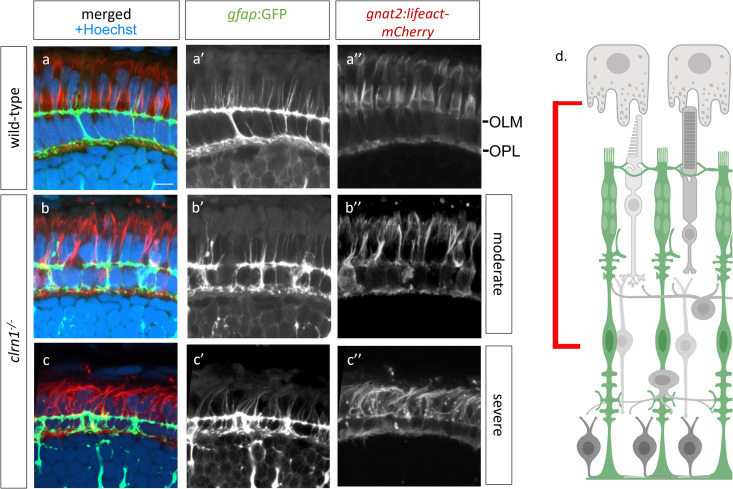
Apical processes of Müller glia and calyceal processes of cone photoreceptors are disorganized in clrn1-/- retina.

Transverse sections from 7 dpf (a) wild-type and (b,c) clrn1-/- mutant zebrafish each expressing the Tg(gfap:GFP); Tg(gnat2:Lifeact-mCherry) transgenes highlighting either (b) moderately or (c) more severely altered Müller glia apical microvilli projections (b’,c’) and cone-specific actin structures (b,” c”) in clrn1-/- mutants. (d) Cartoon showing the area imaged (red bar). Scale bar = 10 µm. OLM, Outer limiting membrane; OPL, Outer plexiform layer. Schematic in (d) created in BioRender under License: https://BioRender.com/x12n534.

Acknowledgments
This image is the copyrighted work of the attributed author or publisher, and ZFIN has permission only to display this image to its users. Additional permissions should be obtained from the applicable author or publisher of the image. Full text @ PLoS Genet.