IMAGE

Figure S1.

ID
ZDB-IMAGE-250320-2
Source
Figures for Doan et al., 2025
Image
Figure Caption

Figure S1.

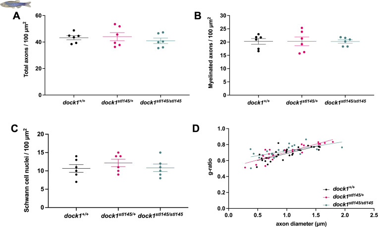
dock1 MUT zebrafish do not exhibit myelin defects at 4 mo. (A–D) Quantifications of the total number of axons, the number of myelinated axons, the number of SC nuclei, and g-ratio were obtained from analyzing TEM micrographs (see Fig. 1). None of these analyses revealed significant differences between WT, HET, or homozygous MUT zebrafish. n = 6 fish per genotype. (A–C) One-way ANOVA with Brown–Forsythe test.

Acknowledgments
This image is the copyrighted work of the attributed author or publisher, and ZFIN has permission only to display this image to its users. Additional permissions should be obtained from the applicable author or publisher of the image. Full text @ J. Cell Biol.