IMAGE

Fig. 1.

ID
ZDB-IMAGE-250219-47
Source
Figures for Gjorcheska et al., 2025
Image
Figure Caption

Fig. 1.

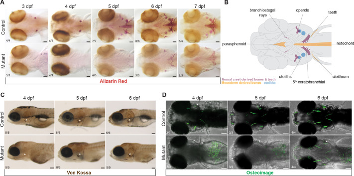
Mineralization deficit in zebrafish sox10 mutants. (A) Example images illustrating a major delay in initiation of bone mineralization in sox10 mutants between 3 and 7 dpf revealed by Alizarin Red staining. Some mineralization is present by 5 dpf but never achieves control levels before lethality at 8 dpf. (B) Schematic representation of the affected mineralized structures and their embryonic origins. (C,D) Von Kossa (C) and OsteoImage (D) staining showing absent calcium deposition and hydroxyapatite formation in sox10 mutants at 4 dpf and gradual recovery starting at 5 dpf. Arrowheads point to the opercle (op). Numbers in panels indicate the proportion of larvae of that genotype with the presented phenotype. Scale bars: 100 µm.

Figure Data
Acknowledgments
This image is the copyrighted work of the attributed author or publisher, and ZFIN has permission only to display this image to its users. Additional permissions should be obtained from the applicable author or publisher of the image. Full text @ Development