
|
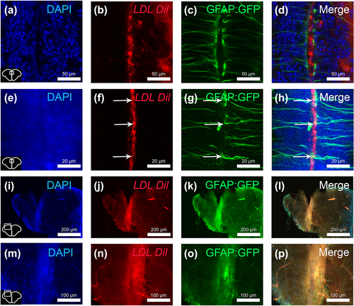
Fig. 8 LDL are not taken up by neural stem cells in homeostatic and regenerative conditions. (a–h) Intracerebroventricular injection of Dil-labelled LDLs in Tg (GFAP::GFP) fish allowing to show that LDLs (in red) are not widely taken up by neural stem cells in homeostatic conditions. Arrows show little LDL detection in GFP positive neural stem cells. (i–p) Intraventricular injection of fluorescent LDLs followed by stab wound injury allows the diffusion of LDL particles within the brain parenchyma but not their increased uptake by neural stem cells. In this context, LDLs were intraperitoneally injected 30 min prior the stab wound and euthanized 1 h30 after this injury. (m–p) Magnification of above pictures (i–k). Bar: 200 μm (i–l), 100 μm (m–p), 50 μm (a–d) and 20 μm (e–h).