
|
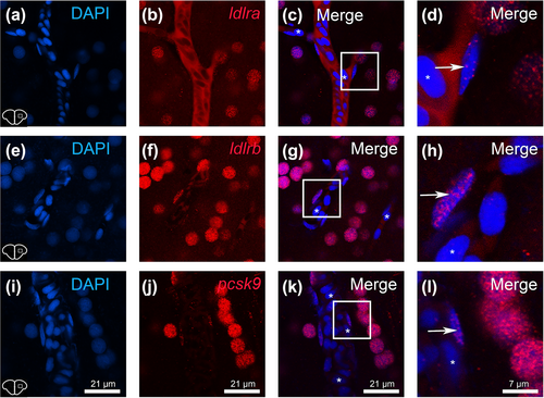
Fig. 6 ldlra, ldlrb and pcsk9 gene expression in endothelial cells. (a–l) Fluorescence in situ hybridization (red) for ldlra, ldlrb and pcsk9 (B, F and J) in the telencephalon of adult zebrafish with DAPI counterstaining (blue, a,e,i). (c,g,k,d,h,l) Merged images with high magnifications of the corresponding white squares. The arrows indicate the detection of transcripts in some cells exhibiting an elongated and flat nucleus localized along the blood vessels, corresponding to endothelial cells. Note that a weak expression is shown in endothelial cells for ldlra and pcsk9, whereas ldlrb appears qualitatively more expressed in these cells. Asterisks show DAPI nuclei without ISH staining. Bar: 21 μm (a,b,c,e,f,g,i,j,k) and 7 μm (d,h,l).