IMAGE

Figure 3

ID
ZDB-IMAGE-250109-351
Source
Figures for Marchica et al., 2024
Image
Figure Caption

Figure 3

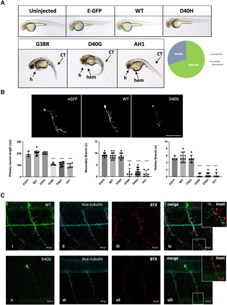
Misexpression of p.D40G, p.G38R and AH1-Annexin A11 in AB Zebrafish result in severe morphological defects at 48 hpf. (A) Lateral view of AB larvae injected at the one-cell stage expressing MNX1:GAL4 and a mix of either empty-UAS GFP, or human Annexin A11-GFP plasmids of wild-type (WT), the non-disease control D40H and mutants D40G, G38R and AH1 (alpha helical 1 deletion) and imaged at 48 hours post-fertilization (hpf). Approximately 30% of larvae with G38R, D40G and AH1 mutants showed a range of morphological phenotypes i.e. curly tail (CT), haemorrhage (hem) and heart defects (h) (n = 40 each group, n = 12 G38R, D40G and AH1 deformed embryos each time, four biological replicates). (B) Live imaging of caudal primary (CaP) motor neurons from normal like GFP positive (+ve) larvae (free of morphological abnormalities) showing representative images (left to right) injected with empty vector-eGFP, wild-type Annexin A11-eGFP and D40G-Annexin A11-eGFP. Neurons imaged from sagittal and cross-sectional view show a single neuron innvervated by a cap neuron (top). Quantitative analysis of axonal length and branching after microinjection with control eGFP vector, wild-type Annexin A11 and amyotrophic lateral sclerosis (ALS) and AH1 mutants (bottom). Statistical analysis was performed on GraphPad Prism 7 using a one-way ANOVA with a post hoc Tukey’s multiple comparisons test. ****P ≤ 0.0001, n = 3: eGFP: n = 12 WT-Annexin A11-eGFP, n = 12: D40G- n = 6 G38R and n = 6 AH1 Annexin A11-eGFP (one axon per biological replicate, maximum projection). Scale bar = 100μm. (C) The D40G mutation results in a reduced amount of axonal Annexin A11 expression and clustering with acetylcholine receptors. [C(i–viii)] Confocal images of zebrafish trunk, lateral view, anterior to the right, imaged at 48 hours post-fertilization (hpf) after microinjection with WT-Annexin A11-eGFP and D40G-Annexin A11-eGFP mutant plasmids and followed by GFP [C(i and v)], ac-Tubulin [C(ii and vi, cyan)] and staining BTX [C(iii and vii, red)]. GFP immunohistochemistry (IHC) demonstrates decreased or absent branching of motor axons in D40G injected embryos, compared to embryos injected with WT-Annexin A11. Insets on [C(iv and viii)] represent effects on postsynaptic development (red arrows and white square). Max projections. Scale bar = 100μm. eGFP = enhanced green fluorescent protein.

Acknowledgments
This image is the copyrighted work of the attributed author or publisher, and ZFIN has permission only to display this image to its users. Additional permissions should be obtained from the applicable author or publisher of the image. Full text @ Brain